Glaucoma is an important cause of permanent vision loss, yet it often develops without pain or obvious early symptoms. For families in Aizawl and across Mizoram, this makes regular eye examinations especially valuable. A person may feel that their eyesight is normal while changes are already occurring in the optic nerve, the structure that carries visual information from the eye to the brain.
This patient guide explains glaucoma screening in Aizawl, including symptoms, risk factors, eye-pressure testing, optic-nerve assessment, visual-field testing and available treatments. It is intended to help patients prepare for a consultation with Dr. Lynapawngia Solo and the eye-care team at Dr. Nunu Eye Hospital & Research Centre. It is general education, not a diagnosis or an individual treatment plan.
What is glaucoma?
Glaucoma is the name for a group of eye diseases that damage the optic nerve. The optic nerve contains more than a million nerve fibres and is essential for vision. In many people, glaucoma is associated with pressure inside the eye that is too high for that particular optic nerve. However, glaucoma can also occur when a pressure reading is within the statistically normal range.
The eye continuously produces a clear fluid called aqueous humour. This fluid normally leaves the eye through drainage tissues near the angle between the cornea and iris. When drainage becomes inefficient or blocked, pressure can rise. Over time, pressure and other factors may damage optic-nerve fibres. The resulting vision loss cannot usually be restored, which is why detection before advanced damage is so important.
Why glaucoma screening matters in Aizawl and Mizoram
Most common open-angle glaucoma progresses gradually. Central reading vision may remain clear until later, while peripheral or side vision is affected first. Because both eyes often work together, the stronger areas of one eye can compensate for the other, making early loss difficult to notice during ordinary activities.
People living outside Aizawl may also need to plan travel and follow-up visits. Early assessment can make that planning more manageable. A glaucoma consultation can establish a baseline, identify whether further tests are needed and determine a safe review schedule. Patients already diagnosed with glaucoma should keep monitoring appointments even when they feel well and their glasses remain unchanged.
Common types of glaucoma
Primary open-angle glaucoma is the most familiar form. The drainage angle remains open, but fluid does not drain efficiently enough. Damage usually develops slowly and without early discomfort. Normal-tension glaucoma causes characteristic optic-nerve damage even though measured eye pressure is not unusually high. This is one reason that a pressure check alone cannot rule out glaucoma.
Angle-closure glaucoma occurs when the iris blocks the drainage angle. It may develop slowly, but a sudden complete blockage can cause a rapid rise in pressure and is an emergency. Secondary glaucoma can follow eye inflammation, injury, certain medicines, advanced cataract or other eye conditions. Babies and children can rarely have developmental forms of glaucoma that require specialist care.
Does glaucoma cause symptoms?
Early open-angle glaucoma usually causes no noticeable symptoms. As damage progresses, patches of side vision may be lost. A person may begin bumping into objects, missing steps, finding crowded environments difficult or struggling to notice vehicles and people approaching from the side. Advanced glaucoma can narrow the field of vision substantially.
Symptoms alone are therefore an unreliable way to detect glaucoma. Clear central vision and the absence of pain do not prove that the optic nerve is healthy. Comprehensive glaucoma screening in Aizawl combines several findings rather than relying on one symptom or one pressure measurement.
Emergency symptoms that need immediate attention
Sudden angle closure can cause severe eye pain, a red eye, rapidly blurred vision, coloured halos around lights, headache, nausea or vomiting. These symptoms require urgent medical care. Do not wait for a routine appointment or try to treat the problem with over-the-counter drops. Sudden vision loss, new flashes or floaters, injury and a curtain-like shadow are also urgent symptoms, although they may have causes other than glaucoma.
Who should consider glaucoma screening?
Anyone can develop glaucoma, but risk is higher with increasing age, a parent or sibling with glaucoma, previously elevated eye pressure, thin central corneas, significant short-sightedness or long-sightedness, previous eye injury, eye inflammation or certain eye operations. Diabetes, blood-pressure problems and other health conditions can influence an ophthalmologist’s overall assessment.
Long-term corticosteroid medicines can raise eye pressure in some people. Steroids may be given as eye drops, tablets, inhalers, injections or skin preparations. Never stop a prescribed medicine without speaking to the clinician who prescribed it. Instead, tell your ophthalmologist about every medicine you use so that appropriate monitoring can be arranged.
If glaucoma runs in your family, encourage close relatives to have comprehensive eye examinations. Family members should not assume they are safe simply because they have no symptoms. The timing of reviews depends on age, examination findings and individual risk; your eye-care professional can recommend a suitable interval.
What happens during glaucoma screening in Aizawl?
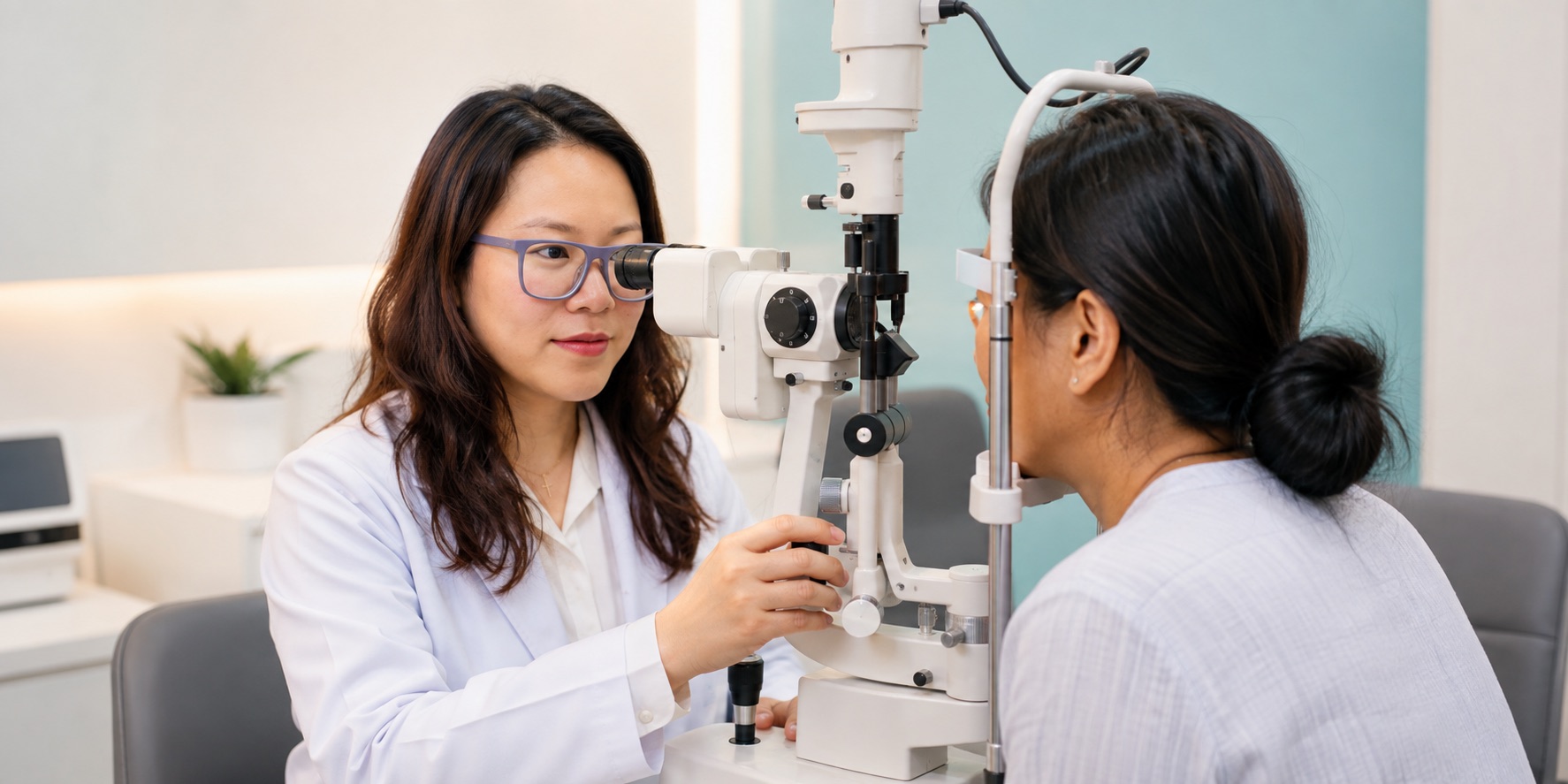
A glaucoma assessment normally begins with questions about vision, previous eye problems, injuries, operations, general health, family history and medicines. Visual acuity is checked, and spectacle power may be measured. The ophthalmologist then examines the front and back of the eyes using a slit lamp and other instruments.
Dilating drops may be used to widen the pupil so the optic nerve and retina can be examined carefully. The drops can temporarily blur near vision and increase sensitivity to light. Patients travelling through bright daylight may find sunglasses helpful, and some people prefer to arrange transport rather than drive immediately after dilation.
Eye-pressure measurement: tonometry
Tonometry measures intraocular pressure. Depending on the instrument, anaesthetic drops and a small amount of dye may be used, or a non-contact method may deliver a brief puff of air. The test is quick. A single result must be interpreted carefully because eye pressure naturally varies during the day and differs between people.
High pressure increases risk, but it does not automatically mean that glaucoma is present. Some people have ocular hypertension without detectable nerve damage, while others develop glaucoma at lower pressures. Corneal thickness and other anatomical factors can influence the reading. Diagnosis therefore requires a complete picture.
Examining the drainage angle
Gonioscopy helps the ophthalmologist see whether the drainage angle is open, narrow or closed. After numbing drops, a special lens may be placed gently on the eye. Understanding the angle is important because open-angle and angle-closure disease have different risks and may require different treatment approaches.
Optic-nerve examination and OCT imaging
The optic nerve is inspected for its colour, shape and the size of the central cup. Photographs can document its appearance for comparison over time. Optical coherence tomography, commonly called OCT, uses light to measure layers of retinal nerve fibres and the optic-nerve region. It is non-invasive and generally takes only a few minutes.
OCT is valuable, but no scan should be interpreted in isolation. Image quality, natural anatomical differences, short-sightedness and other retinal or optic-nerve conditions can affect results. The clinician compares scans with the physical examination, pressure history and visual-field findings.
Visual-field testing
A visual-field test maps peripheral and central sensitivity. You look at a central target and press a button when small lights appear. The lights vary in brightness and location. The test does not hurt, but concentration is important. It is normal to miss some lights, and you should not move your eyes around trying to search for them.
Learning the test can take practice, so an unusual first result may need to be repeated. Reliable follow-up fields help determine whether vision is stable or changing. Try to rest beforehand, bring your reading spectacles if requested and ask for a pause if you become tired.
How is glaucoma diagnosed?
There is no single test that answers every glaucoma question. Diagnosis is based on the relationship between optic-nerve appearance, retinal nerve-fibre measurements, visual-field results, eye pressure, corneal thickness, drainage-angle anatomy and risk factors. Sometimes the first visit establishes suspicion rather than a definite diagnosis, and repeat measurements show whether change is occurring.
A glaucoma suspect may have elevated pressure, an unusual-looking optic nerve or another risk factor without confirmed functional loss. Monitoring is still important. The goal is to avoid unnecessary treatment while also identifying genuine progression early enough to protect vision.
Glaucoma treatment options
Current glaucoma treatment aims to lower eye pressure and reduce the chance of further optic-nerve damage. It cannot normally restore nerve tissue or vision already lost. The target pressure is individualized according to the type and severity of disease, starting pressure, age, life expectancy, health of the other eye and evidence of progression.
Prescription eye drops are a common treatment. Different groups reduce fluid production or improve drainage. Use drops exactly as directed, even when the eye feels normal. Tell the ophthalmologist about breathing problems, heart conditions, allergies, pregnancy and other medicines, because these can influence the safest choice. Report side effects rather than stopping treatment silently.
Laser procedures can help lower pressure in selected forms of glaucoma. Laser trabeculoplasty may improve drainage in open-angle disease, while laser iridotomy creates a small opening in the iris for eyes at risk of angle closure. Surgery may be advised when medicines and laser do not provide sufficient control, or when the disease is advanced. Options include trabeculectomy, drainage implants and several less invasive procedures. Suitability varies from eye to eye.
Using glaucoma eye drops correctly
Wash and dry your hands, tilt your head back and gently pull down the lower eyelid to form a pocket. Place one drop in the pocket without touching the bottle tip to the eye, eyelashes or skin. Close the eye gently; squeezing hard can push medicine out. Pressing lightly at the inner corner beside the nose for one to two minutes may reduce drainage into the throat.
If more than one type of drop is prescribed, allow several minutes between them unless instructed otherwise. Keep a written schedule, use phone reminders and carry enough medicine when travelling from other parts of Mizoram. At each visit, bring your bottles or an accurate list. Ask the clinic to demonstrate again if the technique is difficult.
Living with glaucoma and planning follow-up
Glaucoma care is usually long term. Follow-up may include repeat pressure measurements, optic-nerve examination, OCT and visual fields. A stable pressure reading is encouraging but does not by itself prove that the disease is stable. Keeping appointments allows the clinician to compare results across time and adjust the target pressure when necessary.
Healthy daily habits support general wellbeing but do not replace glaucoma treatment. Avoid smoking, remain physically active as advised by your doctor and manage diabetes and blood pressure. Tell every healthcare professional that you have glaucoma. Before using steroid medicines or over-the-counter eye drops, ask whether they are appropriate for you.
Frequently asked questions
Can glaucoma be cured?
Glaucoma cannot currently be cured, and lost vision is generally permanent. Effective treatment and monitoring can often slow or stop additional damage, especially when disease is identified early.
Is glaucoma the same as high eye pressure?
No. High pressure is an important risk factor, but diagnosis depends on the optic nerve and visual function. Some people with high pressure never develop damage, while normal-tension glaucoma occurs without unusually high readings.
Will new spectacles treat glaucoma?
Spectacles correct refractive blur but do not lower pressure or prevent optic-nerve damage. A person can need both an updated spectacle prescription and glaucoma treatment.
Should relatives be examined?
Yes. A family history increases risk. Parents, siblings and adult children should tell their eye-care professional about the diagnosis and ask when comprehensive screening is appropriate.
Arrange glaucoma screening in Aizawl
If you have a family history of glaucoma, raised eye pressure, increasing age, long-term steroid use or another risk factor, consider arranging a comprehensive examination at Dr. Nunu Eye Hospital & Research Centre in Aizawl, Mizoram. Dr. Lynapawngia Solo can review your history, examine the optic nerves and explain whether observation, further testing or treatment is appropriate.
Patients travelling from other parts of Mizoram can ask the hospital what tests may be required and whether follow-up visits can be coordinated. Bring previous prescriptions, test reports and all current eye drops. Early, consistent care offers the best opportunity to preserve useful vision.
Reliable glaucoma information
For further general reading, see the National Eye Institute overview of glaucoma, its guidance on glaucoma and eye pressure, and the World Health Organization fact sheet on vision impairment.
This article is for general education only. It does not diagnose glaucoma or replace an examination by a qualified ophthalmologist. Seek urgent medical care for severe eye pain, a red eye with nausea or vomiting, sudden vision loss, new flashes or floaters, eye injury or a curtain-like shadow across your sight.