Clear vision. Compassionate care. Better tomorrow.Call: +91 98620 19216
☎ Call now

Author: Dr. Lynapawngia Solo

  • Eye Pain and Headache in Aizawl: Common Causes and Warning Signs

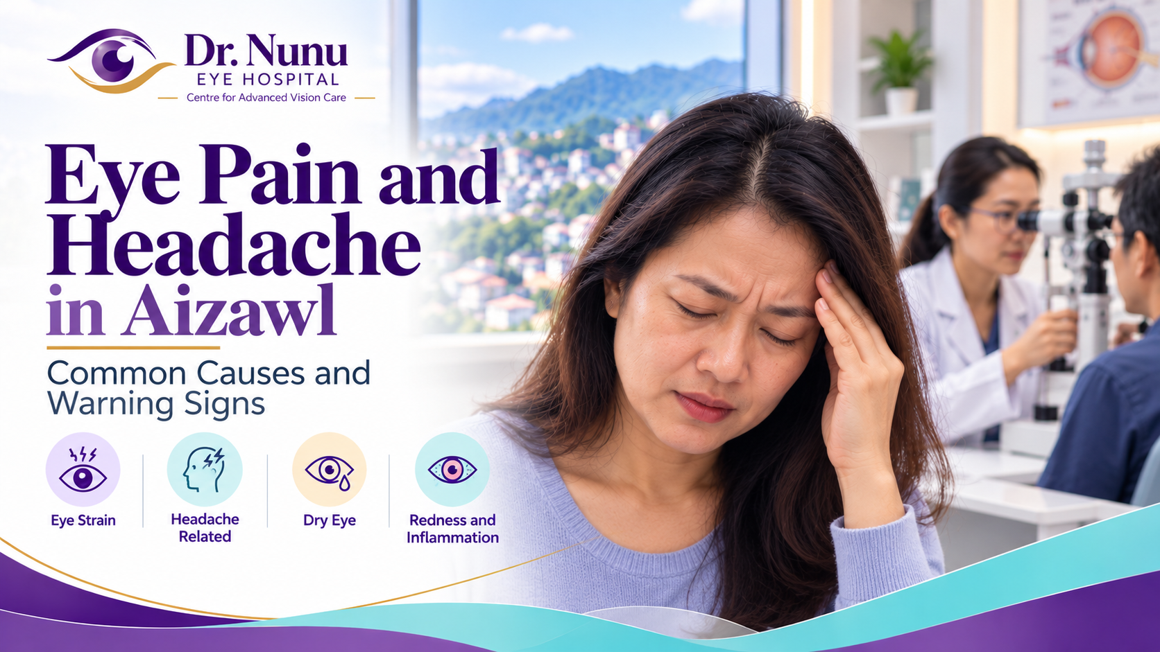
    Eye Pain and Headache in Aizawl: Common Causes and Warning Signs

    Eye pain and headache in Aizawl are common complaints, but the cause is not always simply “eye strain” or a change in spectacle power. Discomfort may come from the surface of the eye, the structures inside or around it, a headache disorder such as migraine, sinus inflammation or, occasionally, an eye or neurological emergency.

    The pattern of symptoms matters. Mild tiredness after prolonged screen use is very different from sudden severe pain with a red eye and blurred vision. This guide explains common causes, warning signs and the eye tests that may help patients in Aizawl and across Mizoram understand when to arrange a routine examination and when to seek urgent medical attention.

    Can an eye problem cause a headache?

    Yes. The eyes, forehead, sinuses and several parts of the brain share sensory nerve pathways. Problems affecting the cornea, eye muscles, focusing system, pressure inside the eye or tissues around the eye can produce pain felt in the forehead, brow or temple. However, many headaches felt “behind the eyes” originate outside the eye. Migraine, tension-type headache, sinus disease and some neurological conditions may all be perceived as eye pain.

    Location alone cannot identify the cause. An examination is particularly important when pain is new, recurrent, one-sided, severe, associated with visual change or different from a person’s usual headache.

    Common causes of eye pain and headache

    Digital eye strain

    Long periods on phones, laptops or other screens can cause tired eyes, brow ache, fluctuating blur, burning and difficulty refocusing. People blink less frequently while concentrating, which destabilises the tear film. Glare, poor lighting, small text, an uncomfortable viewing distance and an uncorrected prescription can make symptoms worse.

    Short breaks, intentional blinking, appropriate lighting and a comfortable screen position may help. The 20-20-20 habit—looking about 20 feet away for 20 seconds every 20 minutes—is a useful reminder, but persistent pain or blur still deserves an eye examination.

    Uncorrected spectacle power

    Myopia, hyperopia and astigmatism may make the visual system work harder. Hyperopia can be especially tiring during reading, while astigmatism can cause blur and squinting at several distances. Presbyopia usually becomes noticeable after about age 40 and may cause near-work fatigue when reading correction is inadequate.

    A refraction test can determine whether a new prescription is needed. Spectacles should not be changed solely because of headache without checking the health of the eyes and considering non-eye causes.

    Dry eye disease

    Dry eye can cause burning, grittiness, heaviness, light sensitivity and fluctuating vision. Wind, fans, air conditioning, smoke, dust, dehydration, reduced blinking, contact lenses and some medicines may aggravate symptoms. Discomfort may extend around the eyes and contribute to a headache.

    Treatment depends on the cause and may involve blinking and screen changes, prescribed lubricants, eyelid care or management of meibomian-gland dysfunction. Read our guide to dry-eye treatment in Aizawl and Mizoram.

    Eye allergy

    Allergic conjunctivitis commonly causes intense itching, watering, redness and puffy eyelids. Rubbing may add soreness or headache. Itching is often more prominent than true deep pain. Significant pain, light sensitivity or reduced vision is not typical of a simple allergy and should prompt an examination.

    Patients with seasonal or recurrent symptoms may find our article on eye-allergy treatment in Aizawl helpful.

    Blepharitis and eyelid-gland problems

    Inflammation around the eyelashes and blocked eyelid oil glands can cause burning, crusting, tenderness and variable blur. A stye or chalazion may produce a localised painful lump. Warm compresses may be advised for some eyelid conditions, but squeezing a lump can worsen inflammation. Recurrent or unusual eyelid swelling needs professional review.

    Contact-lens irritation or infection

    A poorly fitting, damaged, dirty or overworn contact lens can irritate the cornea. More serious contact-lens-related keratitis can cause increasing pain, redness, watering, light sensitivity and reduced vision. Remove the lens and seek prompt eye care when pain or vision change persists. Do not sleep, shower or swim in lenses unless the prescribing professional has specifically approved that use.

    Corneal abrasion or foreign body

    A scratch, dust particle, plant material or other foreign body can cause sharp pain, tearing, blinking discomfort and sensitivity to light. Metal work, drilling and grinding can send high-speed particles into the eye, sometimes without an obvious wound. Chemical exposure requires immediate, prolonged rinsing with clean running water followed by urgent medical assessment. Do not attempt to remove an embedded object.

    Corneal ulcer or keratitis

    An infection or inflammation of the cornea can threaten sight. Warning features include moderate to severe pain, intense redness, light sensitivity, discharge, a white spot on the cornea and reduced vision. Risk is higher with contact-lens misuse, injury involving soil or vegetation, previous corneal disease and reduced immunity. Urgent diagnosis is essential because treatment depends on the organism and cause.

    See our detailed guide to corneal ulcer and eye-infection treatment in Aizawl.

    Uveitis

    Uveitis is inflammation inside the eye. It may cause a deep ache, redness around the coloured part of the eye, marked light sensitivity, floaters and blurred vision. Some cases are associated with autoimmune or infectious conditions, while others have no identified systemic cause. Uveitis requires examination and prescription treatment; steroid eye drops should not be self-started.

    Acute angle-closure glaucoma

    Most glaucoma develops without pain, but acute angle closure is different. A sudden rise in eye pressure may cause severe eye or brow pain, a red eye, blurred vision, coloured halos around lights, headache, nausea or vomiting. The National Eye Institute advises immediate medical help for intense eye pain with red eye, blurred vision or nausea. Acute angle closure is an emergency because vision can be permanently damaged.

    Optic neuritis

    Inflammation of the optic nerve may cause pain that worsens when moving the eye, reduced vision and colours appearing washed out. The eye can look externally normal. New loss of vision with pain on eye movement requires urgent assessment and may need neurological investigation.

    Sinus-related pressure

    Inflamed sinuses can produce pressure around the forehead, cheeks and eyes, often with nasal blockage, discharge, facial tenderness or reduced smell. Not every headache around the eyes is sinusitis. Recurrent episodes without nasal symptoms may instead be migraine or another headache disorder. Swelling around an eye, fever, double vision, reduced vision or painful eye movements alongside sinus symptoms needs urgent care.

    Migraine

    Migraine can produce throbbing headache, nausea and sensitivity to light or sound. Some people experience visual aura, such as shimmering zigzags or a spreading blind spot, usually affecting the visual field of both eyes even if it seems stronger on one side. A first episode, a persistent visual disturbance, symptoms confined to one eye, weakness, speech difficulty or an unusually sudden severe headache requires urgent medical assessment rather than self-diagnosis.

    Tension-type headache

    Tension-type headache often feels like pressure or a tight band around the head and may be associated with stress, poor sleep, dehydration, skipped meals, jaw clenching or neck strain. The eyes may feel heavy even when the eyes themselves are healthy. An eye examination can rule out ocular contributors, but persistent headaches may also require evaluation by a physician.

    Cluster headache

    Cluster headache causes extremely severe one-sided pain around or behind an eye, often with tearing, nasal congestion, eyelid droop or restlessness. Attacks may occur in repeated patterns. Because other urgent conditions can cause similar symptoms, a new severe one-sided headache needs prompt medical assessment.

    Warning signs that need urgent treatment

    Seek urgent eye or emergency medical care for any of the following:

    • sudden severe eye pain or a rapidly worsening headache;
    • eye pain with sudden loss, dimming or marked blurring of vision;
    • a painful red eye with nausea, vomiting or coloured halos around lights;
    • new double vision, drooping eyelid or unequal pupils;
    • weakness, numbness, facial droop, confusion, fainting or difficulty speaking;
    • a “worst-ever” headache or a headache reaching maximum intensity suddenly;
    • eye injury, high-speed foreign body or chemical exposure;
    • pain with light sensitivity and a white spot on the cornea;
    • fever with eyelid swelling, restricted eye movement or a bulging eye;
    • new flashes, many floaters or a curtain-like shadow in the vision;
    • new headache after age 50, especially with scalp tenderness, jaw pain while chewing or vision change;
    • headache with fever, stiff neck, seizure or altered consciousness.

    Do not wait for a routine appointment when these symptoms occur. Our emergency eye-care guide for Aizawl explains additional symptoms that need immediate attention.

    When should you book an eye examination?

    Arrange an examination when eye pain or headache is recurrent, continues despite rest, interferes with reading or work, or is accompanied by blur, redness, light sensitivity or contact-lens discomfort. Children who frequently rub their eyes, close one eye, sit unusually close to screens or complain of headaches after schoolwork should also be assessed.

    A normal eye examination is still useful: it can exclude important ocular causes and help the patient and physician focus on migraine, sinus, neurological or general-health factors where appropriate.

    How an ophthalmologist investigates eye pain

    Symptom and medical history

    The doctor will ask when the pain began, whether it affects one or both eyes, what it feels like, how long it lasts and what triggers or relieves it. Bring information about vision changes, nausea, fever, trauma, contact lenses, medicines, migraine, diabetes, blood pressure and previous eye problems.

    Visual acuity and refraction

    Each eye is tested separately. Refraction identifies myopia, hyperopia, astigmatism and near-vision needs, but a spectacle test is only one part of a pain assessment.

    Slit-lamp examination

    A microscope allows close examination of the eyelids, tear film, cornea, anterior chamber, iris and lens. Fluorescein dye may reveal an abrasion, dry patches, ulcer or contact-lens injury.

    Eye-pressure and angle assessment

    Eye pressure may be measured, and the drainage angle assessed when glaucoma is suspected. A pressure reading must be interpreted together with symptoms, corneal findings and optic-nerve appearance.

    Pupil, eye movement and optic-nerve tests

    Pupil reactions, colour vision, visual fields and eye movements help assess the optic nerves and neurological pathways. Dilating drops may be used to examine the retina and optic nerve. Imaging or referral for medical or neurological evaluation may be recommended when the findings point beyond the eye.

    Safe steps while arranging care

    • Rest from close work and reduce glare if symptoms are mild and clearly linked to prolonged screen use.
    • Remove contact lenses when an eye is painful or red, and do not reinsert them until advised.
    • Do not use someone else’s eye drops or leftover antibiotic or steroid drops.
    • Do not rub a painful eye or attempt to remove an embedded object.
    • After chemical exposure, rinse continuously with clean running water and seek emergency care.
    • Do not drive when vision is blurred, double or otherwise unsafe.

    Painkillers may temporarily reduce symptoms but can also delay recognition of a serious problem. Repeated use of headache medicine can itself contribute to medication-overuse headache, so frequent symptoms need diagnosis rather than continued self-treatment.

    Frequently asked questions

    Can weak eyesight cause headaches every day?

    An uncorrected prescription may contribute to fatigue and headache, especially during near work, but daily headaches should not automatically be blamed on spectacle power. Eye disease, migraine, sleep, hydration, blood pressure, medicines and other medical causes may need consideration.

    Does high eye pressure always cause pain?

    No. Common open-angle glaucoma is usually painless and may have no early symptoms. Sudden angle closure can cause severe pain, redness, blurred vision, halos and nausea and requires emergency treatment.

    Why do my eyes hurt after using a phone?

    Reduced blinking, dry eye, small text, glare, prolonged focusing and an uncorrected prescription can all contribute. Regular breaks and better viewing habits may help, but persistent or one-sided pain, redness or vision change needs examination.

    Can migraine affect vision?

    Yes. Migraine aura can cause temporary visual patterns or blind areas. However, first-time visual symptoms, loss of vision in one eye, neurological symptoms or an abrupt severe headache require urgent medical assessment.

    Is a painful red eye always conjunctivitis?

    No. Conjunctivitis often causes irritation or grittiness, but significant pain, light sensitivity or reduced vision can indicate corneal disease, uveitis or glaucoma. The American Academy of Ophthalmology recommends prompt eye care for moderate to severe pain, blurred vision, light sensitivity or intense redness.

    Eye-pain assessment in Aizawl

    Because eye pain and headache have many possible causes, an accurate diagnosis should come before treatment. Dr. Lynapawngia Solo at Dr. Nunu Eye Hospital & Research Centre can assess vision, the ocular surface, eye pressure, inflammation, optic-nerve and retinal findings, and advise whether additional medical or neurological evaluation is needed.

    To enquire about an eye-pain examination in Aizawl, call +91 98620 19216. If pain is sudden or severe, vision has changed, or neurological symptoms are present, seek emergency medical care without delay.

    Medical note: This article is for general education and cannot diagnose the cause of eye pain or headache. Symptoms can overlap between minor and sight- or life-threatening conditions. Urgent warning signs require immediate professional assessment.

  • LASIK Eye Surgery in Aizawl: Eligibility, Eye Tests, Cost Factors and Recovery

    LASIK Eye Surgery in Aizawl: Eligibility, Eye Tests, Cost Factors and Recovery

    LASIK eye surgery in Aizawl is often considered by adults who want to reduce their dependence on spectacles or contact lenses. LASIK reshapes the clear front surface of the eye—the cornea—so that light focuses more accurately on the retina. It may correct short-sightedness, long-sightedness and astigmatism in appropriately selected patients.

    However, LASIK is not suitable for everyone, and it cannot guarantee perfect vision or freedom from glasses for life. A safe decision begins with a comprehensive eye examination, accurate corneal measurements and a realistic discussion of benefits, alternatives and risks. This guide explains candidacy, preoperative testing, cost factors, recovery and the questions patients in Aizawl and across Mizoram should ask before choosing laser vision correction.

    What is LASIK eye surgery?

    LASIK stands for laser-assisted in situ keratomileusis. During the procedure, the surgeon creates a thin flap in the cornea, reshapes the underlying tissue with an excimer laser and then returns the flap to its original position. Changing the corneal curvature alters how light enters the eye and can reduce refractive error.

    LASIK is an elective refractive procedure. Its purpose is usually to reduce the need for spectacles or contact lenses rather than to treat an eye disease. The National Eye Institute explains that refractive surgery changes the corneal shape and that LASIK works best when the spectacle prescription has remained stable.

    Which vision problems can LASIK correct?

    Myopia or short-sightedness

    In myopia, distant objects look blurred because light focuses in front of the retina. LASIK can flatten the central cornea to move the focus backward toward the retina.

    Hyperopia or long-sightedness

    Hyperopia may cause difficulty with near work and, at higher levels or with age, distance blur. LASIK can steepen the central cornea in selected patients. Eligibility depends on the amount of hyperopia, corneal shape, age and other measurements.

    Astigmatism

    Astigmatism occurs when the cornea or lens has unequal curvature in different directions. Laser treatment may make corneal curvature more regular, but the amount and type of astigmatism influence whether LASIK is appropriate.

    Presbyopia after age 40

    LASIK does not stop presbyopia, the age-related loss of near focusing. A patient who achieves clear distance vision may still need reading glasses. Some people consider monovision, where one eye is targeted for distance and the other for near vision, but not everyone adapts comfortably. A contact-lens trial is often useful before choosing permanent monovision correction.

    Who may be eligible for LASIK?

    Final eligibility can be decided only after examination. Common requirements include:

    • being at least 18 years old, with many surgeons preferring patients over 21;
    • a spectacle or contact-lens prescription that has been stable for at least the period recommended by the surgeon;
    • a refractive error within the safe treatment range of the laser platform;
    • adequate corneal thickness and a regular, healthy corneal shape;
    • healthy tear film and manageable dry-eye symptoms;
    • no active eye infection or uncontrolled eye disease;
    • general health compatible with normal healing;
    • realistic expectations about visual results and possible need for glasses.

    The American Academy of Ophthalmology notes that candidates need healthy, sufficiently thick corneas, generally good eye health and a prescription that has not changed significantly.

    Who may not be suitable for LASIK?

    LASIK may be delayed, discouraged or replaced by another option when a patient has:

    • an unstable spectacle prescription;
    • corneas that are thin, irregular or suspicious for keratoconus;
    • significant untreated dry eye;
    • active infection, inflammation or recurrent corneal herpes;
    • a visually significant cataract;
    • advanced or uncontrolled glaucoma;
    • certain retinal problems;
    • very high refractive error outside a safe corneal treatment range;
    • poorly controlled diabetes or a condition that affects wound healing;
    • pregnancy or breastfeeding, when vision and corneal measurements may fluctuate;
    • medicines that may affect healing or cause unstable vision;
    • unrealistic expectations or an occupation that restricts refractive surgery.

    The U.S. Food and Drug Administration’s LASIK candidacy guidance highlights refractive instability, pregnancy, healing disorders, certain medicines and some occupations or contact sports as issues that require careful discussion.

    Why a detailed LASIK evaluation matters

    A brief spectacle-power test is not enough to determine LASIK safety. Two people with the same prescription may have very different corneal thickness, topography, tear quality, pupil size or retinal health. Preoperative testing is designed to answer two questions: can the refractive error be treated effectively, and can enough structurally healthy cornea be preserved?

    The measurements also guide the laser treatment plan. Contact lenses can temporarily alter corneal shape, so patients are usually asked to stop wearing them before the evaluation for a period based on lens type and the surgeon’s instructions.

    Eye tests before LASIK surgery in Aizawl

    Medical and eye-history review

    The doctor asks about prescription changes, contact-lens use, dry-eye symptoms, previous infection, injury, surgery and family eye history. Diabetes, autoimmune disease, pregnancy, allergies and current medicines are relevant because they may affect measurements or healing.

    Distance and near visual acuity

    Each eye is tested separately with and without correction. This establishes current visual function and helps compare the expected benefit of surgery with vision obtained using glasses.

    Manifest and cycloplegic refraction

    Refraction measures the degree of myopia, hyperopia and astigmatism. In some patients, dilating or cycloplegic drops are used to relax focusing and reveal the full prescription. Stable, repeatable measurements are essential for planning.

    Corneal topography and tomography

    Topography maps the front corneal surface. Tomography assesses the front and back surfaces and corneal thickness distribution. These tests help identify irregular astigmatism, keratoconus or patterns that could increase the risk of corneal weakening after surgery.

    Corneal thickness measurement

    Pachymetry measures corneal thickness. The surgeon calculates how much tissue the laser would remove and how much supporting cornea would remain. A cornea can be clear and appear healthy during a routine examination yet still be unsuitable for LASIK because of its thickness or shape.

    Tear-film and dry-eye assessment

    LASIK can temporarily worsen dry-eye symptoms. The examination may include tear-film stability, corneal staining, eyelid-gland assessment and symptom review. Treating dry eye and blepharitis before surgery can improve comfort and measurement reliability.

    Patients with burning, fluctuating vision or chronic irritation can read our guide to dry-eye treatment in Aizawl and Mizoram.

    Pupil-size measurement

    Pupil size, especially in dim light, may influence the discussion about night glare, halos and treatment-zone planning. It is only one part of risk assessment and must be interpreted with corneal and refractive measurements.

    Slit-lamp examination

    The ophthalmologist examines the eyelids, tear film, cornea, anterior chamber, iris and lens. Active allergy, infection, inflammation, corneal scars or cataract may require treatment or a different plan.

    Eye pressure, optic nerve and retina

    Eye pressure and optic-nerve assessment help screen for glaucoma. A dilated examination may be needed to evaluate the lens and retina, especially in patients with high myopia, diabetes, flashes, floaters or previous retinal disease.

    Preparing for a LASIK consultation

    Bring your current and older spectacle prescriptions if available. Changes over time help establish stability. Bring a list of medicines, details of previous eye problems and information about your work, driving and visual priorities.

    Ask the clinic how long to stop wearing contact lenses before testing. The FDA notes that contact lenses can alter corneal shape for days or weeks depending on the lens type, making measurements inaccurate if they are removed too close to the evaluation.

    Avoid eye makeup on the day of detailed testing if instructed. Tell the doctor about pregnancy, breastfeeding, diabetes, autoimmune disease, severe allergies, eye rubbing and participation in contact sports.

    What happens during LASIK?

    The eye is numbed with anaesthetic drops. A device helps keep the eyelids open. The surgeon creates a thin corneal flap using a femtosecond laser or, in some systems, a microkeratome. The flap is lifted and an excimer laser removes a precisely calculated amount of corneal tissue. The flap is then returned to position, where it adheres without stitches.

    The laser treatment itself is brief, but preparation, positioning and checks take additional time. Patients remain awake and follow instructions to look at a fixation light. Modern systems may track small eye movements, but cooperation remains important.

    Does LASIK hurt?

    Anaesthetic drops reduce pain during the procedure, although pressure or unusual sensations may be noticed. For several hours afterward, eyes can feel gritty, watery, burning or sensitive to light. Patients are normally advised to rest, avoid rubbing and use prescribed drops exactly as directed.

    LASIK recovery: what to expect

    The first day

    Vision may be hazy immediately after surgery and often improves quickly. Arrange for someone to take you home; do not drive on the day of surgery. Protective shields may be provided to prevent accidental rubbing while sleeping.

    The first week

    Use antibiotic, anti-inflammatory and lubricating drops according to the surgeon’s schedule. Attend the early follow-up appointment even if vision feels good. Avoid eye rubbing, makeup, dusty environments and water entering the eyes until cleared.

    Return to work and normal activities

    Many people return to desk work relatively quickly, but timing varies with healing, symptoms and job demands. Driving should resume only after the surgeon confirms vision meets the required standard and the patient feels safe. Swimming, contact sports, heavy exercise and exposure to contaminated water may need to be avoided for longer.

    Vision stabilisation

    Fluctuation, glare, halos and dryness can occur during early recovery. Final healing and visual stability may take weeks to several months. The National Eye Institute notes that the eye may take two to three months to finish healing and that follow-up visits are important.

    Possible benefits of LASIK

    • reduced dependence on spectacles or contact lenses;
    • greater convenience for sports and daily activities;
    • rapid visual recovery for many suitable patients;
    • correction of myopia, hyperopia or astigmatism within appropriate ranges.

    Results vary. Some patients still need glasses for night driving, detailed tasks or residual refractive error. Reading glasses are commonly needed with age because LASIK does not prevent presbyopia.

    Risks and side effects to discuss

    LASIK is surgery and therefore has risks. Possible effects include:

    • dryness, burning or fluctuating vision;
    • glare, halos, starbursts or reduced night-vision quality;
    • under-correction, over-correction or residual astigmatism;
    • continued need for spectacles;
    • infection or inflammation;
    • flap displacement, folds or related complications;
    • corneal haze or delayed healing;
    • corneal weakening or ectasia in susceptible eyes;
    • reduced contrast sensitivity, double vision or persistent visual symptoms;
    • rare loss of best-corrected vision.

    The FDA’s LASIK risk information emphasizes that some patients experience glare, halos, double vision or difficulty seeing in low contrast even when their eye-chart result is good. Personal priorities—especially night driving and occupation—should be part of informed consent.

    Cost factors for LASIK eye surgery in Aizawl

    LASIK cost varies because “laser eye surgery” is not a single identical package. Patients should request a written explanation of what is included rather than comparing only an advertised headline price.

    Type of procedure and technology

    Conventional LASIK, wavefront-guided or topography-guided treatment, femtosecond flap creation and alternative procedures may have different costs. The most expensive option is not automatically the best; suitability and safety should guide the recommendation.

    Preoperative testing

    Comprehensive refraction, topography, tomography, pachymetry, dry-eye evaluation and retinal assessment may be included or billed separately. Ask which tests are necessary and whether repeat measurements are covered.

    Surgeon and facility fees

    The quoted price may include the surgeon, laser facility, medicines, protective shields and follow-up visits—or these may be separate. Confirm whether the quotation covers one eye or both eyes.

    Follow-up and enhancement policy

    Ask how many postoperative visits are included, how long follow-up continues and what happens if residual power remains. Enhancement surgery is not always advisable and may have eligibility rules, waiting periods or additional costs.

    Travel and time away from work

    Patients travelling from outside Aizawl should account for transport, accommodation and the need to return for follow-up. Do not plan surgery without arranging a safe journey home and access to urgent advice if symptoms develop.

    Insurance and financing

    Refractive surgery is often considered elective and may not be covered by standard insurance. Verify coverage directly with the insurer and clinic. Read financing terms carefully and do not allow a limited-time discount to replace medical decision-making.

    LASIK alternatives

    If LASIK is unsuitable, alternatives may include:

    • spectacles;
    • contact lenses;
    • photorefractive keratectomy (PRK);
    • small-incision lenticule extraction (SMILE), where available and suitable;
    • phakic intraocular lenses for selected higher refractive errors;
    • refractive lens exchange in carefully chosen patients;
    • continued monitoring until prescription stability is established.

    Each option has distinct benefits, recovery patterns and risks. Cataract surgery may be more appropriate than corneal laser treatment when a cataract is the main cause of visual decline.

    Questions to ask before choosing LASIK

    • Is my prescription stable and within a safe treatment range?
    • Are my corneal shape and thickness suitable?
    • Do I have dry eye, keratoconus risk, cataract, glaucoma or retinal disease?
    • What procedure is recommended and why?
    • What result is realistic for my eyes?
    • Could I still need distance or reading glasses?
    • What are my personal risks of glare, halos or dry eye?
    • What does the quoted cost include?
    • Who provides postoperative care and emergency advice?
    • What alternative is safest if LASIK is unsuitable?

    Warning signs after LASIK

    Contact the treating surgeon urgently for worsening pain, increasing redness, sudden vision reduction, discharge, significant light sensitivity, trauma to the eye or a displaced-feeling flap. Do not wait for a routine follow-up when symptoms are severe or rapidly changing.

    For general urgent symptoms, see our article on emergency eye care in Aizawl.

    Frequently asked questions

    Will LASIK remove spectacles permanently?

    LASIK may greatly reduce spectacle dependence, but it cannot guarantee that glasses will never be needed. Eyes can change with age, and most people eventually develop presbyopia and need near correction.

    What is the best age for LASIK?

    There is no single best age. Patients must be adults and have stable vision, healthy corneas and appropriate expectations. Eligibility becomes more complex as presbyopia and cataract risk increase.

    Can LASIK correct very high power?

    Sometimes, but safety depends on prescription, corneal thickness, corneal shape and predicted remaining tissue. A phakic lens or another option may be safer for some high prescriptions.

    Can LASIK be done with dry eye?

    Mild dry eye may be treated and stabilised before reassessment. Significant or uncontrolled dry eye may make LASIK unsuitable because surgery can worsen symptoms.

    When can I use screens after LASIK?

    Follow the surgeon’s instructions. Screens do not normally damage the flap, but reduced blinking can worsen dryness. Resume gradually, take breaks and use prescribed lubricants.

    Can I fly after LASIK?

    Air travel may be possible relatively soon, but cabin dryness and follow-up timing matter. Ask the surgeon before booking travel and make sure postoperative review will not be missed.

    LASIK eligibility consultation in Aizawl

    Anyone considering laser vision correction should begin with a comprehensive examination rather than assuming LASIK is the correct procedure. Dr. Lynapawngia Solo at Dr. Nunu Eye Hospital & Research Centre can assess refractive error, eye health and the need for specialised corneal testing, then discuss appropriate next steps or referral where required.

    To enquire about a LASIK eligibility assessment in Aizawl, call +91 98620 19216. Bring current spectacles, previous prescriptions, contact-lens information and a list of medicines. Ask in advance how long contact lenses should be removed before corneal measurements.

    Medical note: This article provides general education and does not confirm that LASIK is suitable for any individual. Refractive surgery is elective and carries risks. A complete examination and informed discussion with a qualified ophthalmologist are essential before treatment.

  • Stye and Chalazion Treatment in Aizawl: Causes, Home Care and When to Visit an Eye Doctor

    Stye and Chalazion Treatment in Aizawl: Causes, Home Care and When to Visit an Eye Doctor

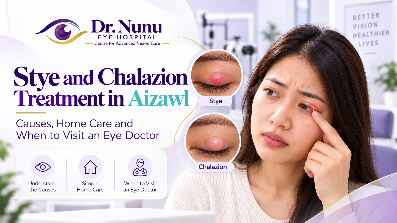
    A painful bump or firm lump on the eyelid is often described simply as a “stye,” but not every eyelid lump is the same. A stye is usually a tender infection near an eyelash or an oil gland, while a chalazion is generally a blocked oil gland that forms a firmer and often less painful swelling. Understanding the difference helps patients choose safe home care and recognise when professional treatment is needed.

    If you are looking for stye and chalazion treatment in Aizawl, this guide explains common causes, warning signs, warm-compress technique, prevention and the circumstances in which an ophthalmologist should examine the eyelid. Most cases improve without surgery, but persistent, recurrent or rapidly worsening lumps should not be ignored.

    What is a stye?

    A stye, medically called a hordeolum, is a localised infection or inflamed blockage involving an eyelash follicle or an eyelid oil gland. An external stye develops near the base of an eyelash and may form a small yellow point. An internal stye forms deeper inside the eyelid and can cause more diffuse tenderness and swelling.

    Typical features include:

    • a painful or tender red lump;
    • localised eyelid swelling;
    • a gritty or irritated feeling;
    • watering of the eye;
    • crusting near the eyelashes;
    • a small pus-filled point in some cases.

    A stye is not the same as general conjunctivitis. Conjunctivitis usually causes widespread redness, watering or discharge without a single tender eyelid lump. Read our guide to conjunctivitis treatment in Aizawl and Mizoram for more information.

    What is a chalazion?

    A chalazion is a lump caused by blockage and inflammation of a meibomian gland, one of the small oil-producing glands inside the eyelid. The trapped oily material triggers inflammation and gradually forms a rounded swelling.

    A chalazion often begins with mild tenderness but usually becomes firm and relatively painless. It can occur on the upper or lower eyelid. A large chalazion may press on the cornea and temporarily blur or distort vision.

    The Government of Mizoram’s Standard Treatment Guidelines for chalazion note that some chalazia resolve spontaneously and that persistent cases may require treatment selected by a clinician.

    Stye versus chalazion: how are they different?

    The two conditions can look similar, particularly in the early stage. The following differences are useful but do not replace an examination:

    • Pain: a stye is commonly sore and tender; a chalazion is often painless after the initial inflammation settles.
    • Location: an external stye usually appears at the eyelash edge; a chalazion frequently feels deeper within the lid.
    • Appearance: a stye may form a small yellow centre; a chalazion is often a smooth, firm lump.
    • Cause: a stye generally involves infection or acute inflammation; a chalazion results mainly from a blocked oil gland.
    • Course: a stye may drain and improve over days; a chalazion may persist for several weeks or longer.

    Some styes evolve into chalazia after the acute infection settles but the gland remains blocked.

    What causes styes and chalazia?

    Blocked eyelid oil glands

    Meibomian glands release oils that help prevent tears from evaporating. Thickened secretions, inflammation or blockage can trap oil within the gland and produce a chalazion.

    Bacteria around the eyelashes

    Styes commonly occur when bacteria enter an eyelash follicle or oil gland. Touching or rubbing the eyes with unwashed hands can transfer bacteria to the eyelid margin.

    Blepharitis

    Blepharitis causes chronic inflammation, crusting and irritation along the eyelid edges. It can interfere with oil-gland function and increase the likelihood of recurrent styes or chalazia.

    Rosacea and oily skin conditions

    People with rosacea, seborrhoeic dermatitis or meibomian gland dysfunction may have thicker gland secretions and repeated eyelid inflammation.

    Eye makeup and contact-lens hygiene

    Sleeping in eye makeup, using expired cosmetics, sharing makeup or handling contact lenses without clean hands can irritate the eyelids and increase infection risk. Makeup applicators may collect oils and bacteria over time.

    Diabetes and other health factors

    Diabetes can increase susceptibility to infections and may affect healing. A clinician may consider general health when eyelid infections are frequent, severe or slow to resolve.

    Safe home care for a stye or chalazion

    Use a warm compress correctly

    Warm compresses are the main home treatment for uncomplicated styes and chalazia. Warmth can soften thickened oils and encourage a blocked gland to open naturally.

    1. Wash your hands thoroughly.
    2. Soak a clean washcloth in comfortably warm—not scalding—water.
    3. Wring out excess water and place the cloth over the closed eyelid.
    4. Keep it in place for about 10 minutes, reheating the cloth as it cools.
    5. Repeat several times a day as advised by your eye doctor.

    The compress should never be hot enough to burn the delicate eyelid skin. Use a fresh, clean cloth and do not share it with another person.

    Gentle eyelid massage

    After warming the lid, a clinician may recommend very gentle massage toward the eyelashes to help gland contents drain. Do not massage a severely painful, very red or rapidly spreading infection without medical advice. Never use sharp tools or fingernails on the lump.

    Keep the eyelids clean

    Wash away crusting gently using the eyelid-cleaning method recommended by your eye doctor. People with blepharitis may require a longer-term lid-hygiene routine even after the lump resolves.

    Pause contact lenses and eye makeup

    Avoid contact lenses and eye makeup until the eyelid is comfortable and healed. Continuing to wear lenses can add irritation and handling may spread bacteria. Replace potentially contaminated eye cosmetics rather than reusing them after the infection resolves.

    Do not squeeze or pop the lump

    Squeezing can push infection into surrounding eyelid tissue and increase swelling. Do not pierce the lump, pull out an eyelash or attempt drainage at home. Drainage should be performed only by an appropriately trained clinician using sterile equipment.

    The NHS guidance on styes recommends warm compresses and specifically warns against bursting a stye because doing so can spread infection.

    What should you avoid?

    • Do not apply toothpaste, garlic, turmeric paste, saliva or unverified herbal preparations near the eye.
    • Do not use leftover antibiotic or steroid eye drops.
    • Do not share towels, cosmetics or contact-lens equipment.
    • Do not rub the eyelid repeatedly.
    • Do not continue contact-lens wear when the eye or eyelid is inflamed.
    • Do not delay care if redness or swelling is spreading.

    Eye medicines are not interchangeable. Steroid-containing drops or ointments can worsen some infections and raise eye pressure, so they should be used only after examination and prescription.

    When should you visit an eye doctor in Aizawl?

    Arrange an ophthalmic examination if:

    • the lump does not begin improving with appropriate home care;
    • it persists for several weeks;
    • pain or swelling is significant;
    • the entire eyelid becomes swollen;
    • the eye itself becomes red or painful;
    • there is pus or heavy discharge;
    • vision becomes blurred or the lump presses on the eye;
    • the swelling keeps returning in the same location;
    • multiple lumps develop repeatedly;
    • the patient has diabetes or reduced immunity;
    • a child cannot open the eye comfortably;
    • you are unsure whether the lump is a stye, chalazion or another eyelid condition.

    A recurrent lump in exactly the same place, loss of eyelashes, unusual bleeding, irregular thickening or a lesion that does not heal requires careful examination. Although uncommon, some eyelid tumours can resemble a persistent chalazion. The doctor may recommend biopsy when a lesion is atypical or repeatedly returns.

    Symptoms that need urgent attention

    Seek urgent care if eyelid redness and swelling spread rapidly, the eye becomes difficult to move, vision falls, the eye protrudes, pain is severe, or fever and general illness develop. These features can suggest infection extending beyond a simple stye.

    Sudden vision loss, chemical exposure, penetrating injury or severe eye pain should be treated as an emergency. See our overview of urgent eye symptoms in Aizawl.

    How an eye doctor examines an eyelid lump

    The ophthalmologist will ask when the lump started, whether it is painful, whether similar lumps have occurred and what treatments have been tried. Contact-lens habits, eye makeup, skin conditions, diabetes and symptoms of blepharitis may be relevant.

    The eyelid is examined from the outside and may be gently turned to inspect its inner surface. A slit-lamp microscope helps evaluate the eyelashes, oil-gland openings, conjunctiva, cornea and the rest of the eye. Vision is checked, particularly if the lump is large or close to the visual axis.

    Additional laboratory tests are rarely needed for a straightforward first episode. Recurrent or atypical lesions may need closer assessment or tissue examination.

    Medical treatment for a stye

    Many styes drain and settle with warmth and hygiene. If infection is significant, the ophthalmologist may prescribe an antibiotic ointment, drops or oral medicine depending on the findings. Antibiotics are not automatically required for every lump.

    A very painful internal stye or a lesion that fails to drain may require a small procedure. This should not be attempted at home. Treatment choice depends on the location of the infection, surrounding swelling and the patient’s health.

    Medical treatment for a chalazion

    Continued conservative treatment

    Small chalazia often improve gradually with warm compresses, lid massage and management of underlying blepharitis. Resolution can take weeks, and the lump may become smaller before disappearing.

    Steroid injection

    For selected chalazia, an ophthalmologist may inject corticosteroid into or near the lesion. This can reduce inflammation, but it has potential risks including skin-colour change and is not suitable for every patient or every location.

    Incision and curettage

    A persistent or visually significant chalazion may be drained through a small incision, usually from the inner eyelid surface. Local anaesthetic is commonly used in adults. Children may require different anaesthesia planning depending on age and cooperation.

    The American Academy of Ophthalmology’s patient guidance explains that surgery may be considered when a stye or chalazion affects vision or fails to resolve, while recurrent lesions may require biopsy.

    Styes and chalazia in children

    Children frequently touch or rub their eyes, and they may find warm-compress treatment difficult. A parent should supervise and make sure the cloth is comfortably warm. Never force a child to tolerate excessive heat or firm pressure.

    Seek medical advice if a child has marked swelling, fever, spreading redness, vision complaints, repeated chalazia or a lump that persists. A large chalazion can sometimes change the corneal shape and blur vision, which is important during visual development.

    Why do eyelid lumps keep returning?

    Recurrence may be related to chronic blepharitis, meibomian gland dysfunction, rosacea, incomplete lid hygiene, eye rubbing or reuse of contaminated cosmetics. Treatment may therefore include a preventive eyelid-care routine rather than focusing only on each individual lump.

    An ophthalmologist can look for signs of gland dysfunction and advise on suitable lid cleaning, warm compress frequency and treatment of associated dry eye. Read more about dry-eye symptoms and treatment in Aizawl.

    How to reduce the risk of future styes and chalazia

    • Wash hands before touching the eyes or handling contact lenses.
    • Remove eye makeup completely before sleeping.
    • Replace expired mascara, eyeliner and applicators.
    • Do not share towels, cosmetics or contact-lens accessories.
    • Follow lens-cleaning and replacement instructions.
    • Manage blepharitis and rosacea consistently.
    • Avoid rubbing the eyes.
    • Use warm compresses as advised if oil-gland blockage is recurrent.
    • Keep diabetes and other medical conditions appropriately managed.

    Frequently asked questions

    Is a stye contagious?

    A stye itself is not usually spread like a cold, but bacteria can be transferred through hands, towels or cosmetics. Good hygiene and avoiding shared personal items reduce this risk.

    Can I go to work or school with a stye?

    Many people can continue normal activities if they feel well, but should avoid sharing towels, rubbing the eye, makeup and contact lenses. Significant pain, discharge or spreading swelling warrants medical advice.

    How long does a chalazion take to disappear?

    Some improve over several weeks, while others last longer. Size, duration, gland blockage and underlying lid inflammation affect recovery. A persistent lump should be examined rather than treated indefinitely at home.

    Do antibiotics cure a chalazion?

    A chalazion is primarily inflammatory and caused by blocked oil, so antibiotics do not automatically remove it. They may be prescribed when infection or associated blepharitis is present.

    Can a chalazion affect eyesight?

    A large lesion can press on the cornea and cause temporary blur or astigmatism. Vision usually improves when pressure resolves, but any visual change should be assessed.

    Should I use a hot compress?

    Use comfortably warm—not hot—water. Excess heat can burn eyelid skin. Test the temperature carefully, especially for children or people with reduced skin sensation.

    Can I wear contact lenses during treatment?

    It is safer to stop lens wear until the eyelid and eye have healed and any prescribed treatment is complete. Ask your eye doctor when it is appropriate to restart.

    Stye and chalazion treatment in Aizawl

    Dr. Lynapawngia Solo at Dr. Nunu Eye Hospital & Research Centre can assess eyelid lumps, blepharitis and related eye irritation. The examination helps distinguish a stye or chalazion from other conditions and determines whether continued home care, medication, a procedure or further testing is appropriate.

    To enquire about stye or chalazion treatment in Aizawl, call +91 98620 19216. Mention severe pain, spreading swelling, fever or vision changes when booking so the clinic can advise on urgency.

    Medical note: This article provides general information and does not replace an eye examination. Do not squeeze, pierce or attempt to drain an eyelid lump yourself. Seek urgent care for reduced vision, severe pain, rapidly spreading swelling, fever or difficulty moving the eye.

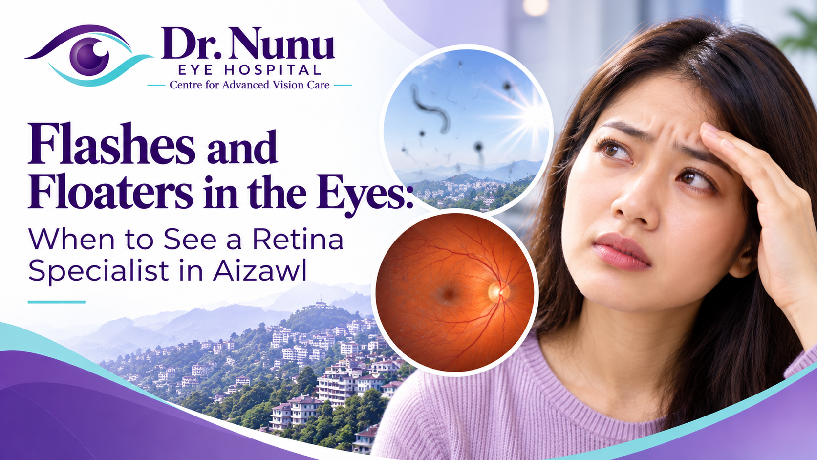
  • Flashes and Floaters in the Eyes: When to See a Retina Specialist in Aizawl

    Flashes and Floaters in the Eyes: When to See a Retina Specialist in Aizawl

    Flashes and floaters in the eyes are common visual symptoms, but a sudden change can sometimes be a warning sign of a retinal tear or retinal detachment. Many people describe floaters as moving dots, threads, cobwebs or small shadows. Flashes may look like brief sparks, lightning streaks or camera flashes at the edge of vision.

    Most long-standing floaters are harmless. However, new symptoms—especially a sudden shower of floaters, repeated flashes, blurred vision or a dark curtain across the sight—need urgent assessment. If you are experiencing these symptoms in Aizawl or elsewhere in Mizoram, do not wait for them to settle on their own. A prompt dilated retinal examination can help protect vision.

    This guide explains why flashes and floaters occur, which symptoms require immediate care, what a retina specialist checks and what treatment may be needed.

    What are eye floaters?

    Floaters are shapes that appear to drift through your field of vision. They may look like:

    • small black or grey dots;
    • threads or wavy lines;
    • rings, circles or specks;
    • cobweb-like shadows;
    • a cluster of tiny spots.

    They often move when the eyes move and seem to slip away when you try to look directly at them. Floaters are usually easier to notice against a bright, plain background such as a white wall, a computer screen or a clear sky.

    The eye is filled with a transparent gel called the vitreous. With age, this gel gradually becomes more liquid and develops tiny clumps or strands. These structures cast shadows on the retina—the light-sensitive tissue at the back of the eye—and the shadows are perceived as floaters.

    What are flashes of light in the eyes?

    Flashes occur when the retina is mechanically stimulated. As the vitreous gel changes or pulls on the retina, a person may see brief streaks or sparks even though no external light is present. They are often more noticeable in a dark room or during eye movement.

    Some people describe flashes as lightning at the side of their vision. Others report flickering, stars or brief arcs of light. Flashes caused by vitreous traction are different from the shimmering zigzag patterns that may accompany migraine, but symptoms can overlap. A first episode or an unusual visual disturbance should be assessed rather than self-diagnosed.

    Why do flashes and floaters happen?

    Normal age-related vitreous changes

    The most common cause is age-related change in the vitreous. As the gel becomes more liquid, it can separate from the retina. This is called a posterior vitreous detachment, or PVD. It becomes more common with age and may cause a sudden increase in floaters and flashes.

    A PVD itself often does not require treatment. The important concern is that the separating vitreous can occasionally pull hard enough to create a retinal tear. Fluid may then pass through the tear and lift the retina away from the wall of the eye, causing a retinal detachment.

    Retinal tear or retinal detachment

    A retinal tear may not cause pain. Its warning signs can be new flashes, many new floaters, a shower of dark dots or a change in vision. If a detachment develops, a shadow or curtain may spread from the side toward the centre of vision.

    The National Eye Institute advises immediate evaluation when many new floaters appear suddenly, especially with flashes, a dark shadow or a blurry area. Retinal tear and detachment are medical emergencies because delayed treatment can lead to permanent vision loss.

    Short-sightedness

    People with moderate or high myopia may develop vitreous changes earlier and can have a higher risk of peripheral retinal weakness, tears or detachment. A person who wears strong minus-powered spectacles should be particularly careful about a sudden change in flashes or floaters.

    Eye surgery or injury

    New floaters after cataract surgery, another eye operation or an eye injury require professional assessment. Trauma can cause bleeding, inflammation or damage to the retina even when the outside of the eye appears normal.

    Inflammation, infection or bleeding

    Inflammation inside the eye, called uveitis, can create floaters and may be accompanied by redness, pain or sensitivity to light. Diabetes and other retinal blood-vessel conditions can cause bleeding into the vitreous, producing dots, haze, cobwebs or a sudden reduction in sight.

    When should you see a retina specialist in Aizawl?

    Seek an urgent eye examination if you notice any of the following:

    • a sudden onset of floaters for the first time;
    • a rapid increase in the number or size of floaters;
    • new flashes of light in one eye;
    • a shower of black dots, specks or cobwebs;
    • a dark curtain, veil or shadow moving across vision;
    • loss of side vision;
    • sudden blurred or reduced vision;
    • flashes or floaters after an eye injury or eye surgery;
    • symptoms accompanied by eye pain or marked redness.

    A dark curtain, sudden loss of vision or rapidly expanding shadow should be treated as an emergency. Contact an ophthalmologist or go to the nearest emergency facility without delay. Do not drive yourself if your vision is impaired.

    For additional warning signs, see our guide to emergency eye care in Aizawl.

    Are long-standing floaters dangerous?

    One or two floaters that have remained unchanged for months or years, without flashes or loss of vision, are commonly harmless. The brain may gradually learn to ignore them, so they become less noticeable. Nevertheless, an eye examination is advisable if they have never been assessed or if you have risk factors such as high myopia, diabetes, previous surgery, trauma or a family history of retinal detachment.

    The key issue is change. Even a person who has had harmless floaters for years should seek prompt care if the number suddenly increases or new flashes, shadows or blurred vision appear.

    Who has a higher risk of retinal tears and detachment?

    Urgent assessment is particularly important for people who:

    • are very short-sighted;
    • are middle-aged or older;
    • have undergone cataract or other eye surgery;
    • have experienced significant eye trauma;
    • have had a retinal tear or detachment in the other eye;
    • have a family history of retinal detachment;
    • have known weak areas in the retina;
    • have diabetes or another retinal blood-vessel disorder.

    The American Academy of Ophthalmology identifies sudden flashes, many new floaters, a side-vision shadow and a grey curtain as early warning signs of retinal detachment that require immediate ophthalmic care.

    What happens during a retina examination?

    Symptom and risk review

    The doctor will ask when the symptoms started, whether they affect one or both eyes and whether they are changing. Describe the exact appearance of flashes and floaters, any missing area of vision, recent injury or surgery and relevant conditions such as diabetes or high myopia.

    Visual-acuity and pupil testing

    Each eye is tested separately. The doctor may also examine pupil reactions because an abnormal response can indicate significant retinal or optic-nerve dysfunction.

    Dilated retinal examination

    Dilating drops widen the pupil so the ophthalmologist can inspect the vitreous, retina and optic nerve. Special lenses and bright illumination are used to examine the central and peripheral retina for tears, holes, bleeding or detachment. Gentle pressure around the eyelid may sometimes be required to view the far retinal periphery.

    Dilating drops can cause temporary glare, light sensitivity and blurred near vision for several hours. Bring sunglasses and consider arranging transport, particularly if your vision is already affected.

    Additional tests

    Optical coherence tomography, or OCT, may be used when the macula needs detailed imaging. Retinal photographs can document findings. If the doctor cannot see the retina clearly because of dense bleeding or another obstruction, an eye ultrasound may be required. The tests selected depend on the examination findings.

    How are flashes and floaters treated?

    Treatment depends entirely on the cause.

    Posterior vitreous detachment without a retinal tear

    If the retina is intact, treatment is often unnecessary. Flashes commonly settle as the vitreous stops pulling, while floaters may persist but become less distracting. The doctor may recommend follow-up because a tear can occasionally develop after the first examination. Return immediately if symptoms worsen.

    Retinal tear

    A retinal tear may be sealed with laser treatment or cryotherapy to create a scar around the break and reduce the chance of fluid passing underneath the retina. The procedure and timing depend on the tear’s location, appearance and associated risk.

    Retinal detachment

    A detached retina usually requires surgery. Options may include pneumatic retinopexy, scleral buckle surgery or vitrectomy. The appropriate method depends on the type and extent of detachment, the position of the retinal breaks and the condition of the lens and vitreous.

    Early treatment generally offers a better opportunity to preserve vision, particularly before the central retina is affected. This is why a curtain-like shadow or sudden visual-field loss must not be ignored.

    Inflammation, bleeding or another retinal condition

    When floaters result from uveitis, infection, diabetic retinal disease or bleeding, the underlying condition must be treated. The ophthalmologist may coordinate care with a physician when diabetes, blood pressure, autoimmune disease or another systemic condition contributes.

    Can eye drops or home remedies remove floaters?

    No routine eye drop or home remedy dissolves age-related vitreous floaters. Hydration, rest or nutritional supplements do not repair a retinal tear. Moving the eyes may temporarily shift a floater away from the centre of vision, but it does not address the cause.

    Do not massage or press the eye, and do not delay an examination while trying home treatments when symptoms are new. Persistent floaters that severely interfere with daily life can occasionally be treated surgically, but vitrectomy carries meaningful risks and is not recommended simply because a small floater is annoying. The risks and benefits require careful discussion with an ophthalmologist.

    Flashes, floaters and migraine: what is the difference?

    Migraine aura often produces shimmering zigzags, coloured patterns or an expanding bright area that affects both eyes, even if it seems stronger on one side. It commonly develops over several minutes and resolves within an hour, sometimes followed by headache.

    Vitreous or retinal flashes are often brief, lightning-like streaks at the edge of vision and may affect one eye. These descriptions are not reliable enough for self-diagnosis. A first episode, a change from your usual migraine pattern or any associated vision loss should be evaluated urgently.

    Practical steps if symptoms begin in Aizawl or elsewhere in Mizoram

    1. Cover one eye at a time. This can help identify which eye is affected, but do not delay care.
    2. Note the time of onset. Tell the doctor whether symptoms appeared suddenly or gradually.
    3. Check for a shadow. Without repeatedly testing or straining, notice whether any part of the side or central vision is missing.
    4. Avoid driving if vision is affected. Ask someone to accompany you.
    5. Seek urgent ophthalmic assessment. Clearly state that you have sudden flashes, new floaters, a curtain or reduced vision.

    People travelling from other parts of Mizoram should not postpone emergency evaluation solely to reach a preferred clinic. If there is sudden vision loss or a curtain-like shadow, attend the nearest facility capable of urgent eye assessment.

    Frequently asked questions

    Do floaters always mean retinal detachment?

    No. Most floaters result from common vitreous changes. However, symptoms alone cannot reliably rule out a retinal tear, particularly when floaters are sudden, numerous or accompanied by flashes or a shadow.

    Is retinal detachment painful?

    It is often painless. The absence of pain does not make sudden flashes, floaters or a curtain safe to ignore.

    Can a routine spectacle test detect a retinal tear?

    A vision or refraction test alone is not enough. The peripheral retina generally needs examination through a dilated pupil by an eye-care professional with appropriate equipment.

    How quickly should new flashes and floaters be checked?

    Sudden symptoms should be assessed urgently, ideally the same day or as directed by an ophthalmologist. A curtain, new field loss or sudden reduction in sight requires immediate emergency care.

    Will floaters disappear?

    They may not physically disappear, but many become less noticeable with time. Persistent symptoms still need reassessment if they suddenly change.

    Can children experience flashes and floaters?

    Yes, although age-related PVD is less common in children. Trauma, inflammation, high myopia and retinal conditions can cause symptoms. A child reporting new flashes, spots or missing vision should be examined promptly.

    Book an urgent retinal examination in Aizawl

    If you notice sudden flashes and floaters in Aizawl, particularly with blurred vision, a shadow or a curtain-like effect, arrange urgent assessment. Dr. Lynapawngia Solo at Dr. Nunu Eye Hospital & Research Centre can evaluate the retina and advise whether monitoring, laser treatment, surgery or referral is required.

    Call +91 98620 19216 and clearly describe sudden or worsening symptoms so the clinic can advise on urgency. If you cannot promptly reach the clinic and vision is suddenly reduced, go to the nearest emergency facility.

    Medical note: This article provides general education and does not replace an examination. Sudden flashes, a shower of new floaters, a dark curtain or sudden vision loss can represent a retinal emergency. Seek urgent professional care.

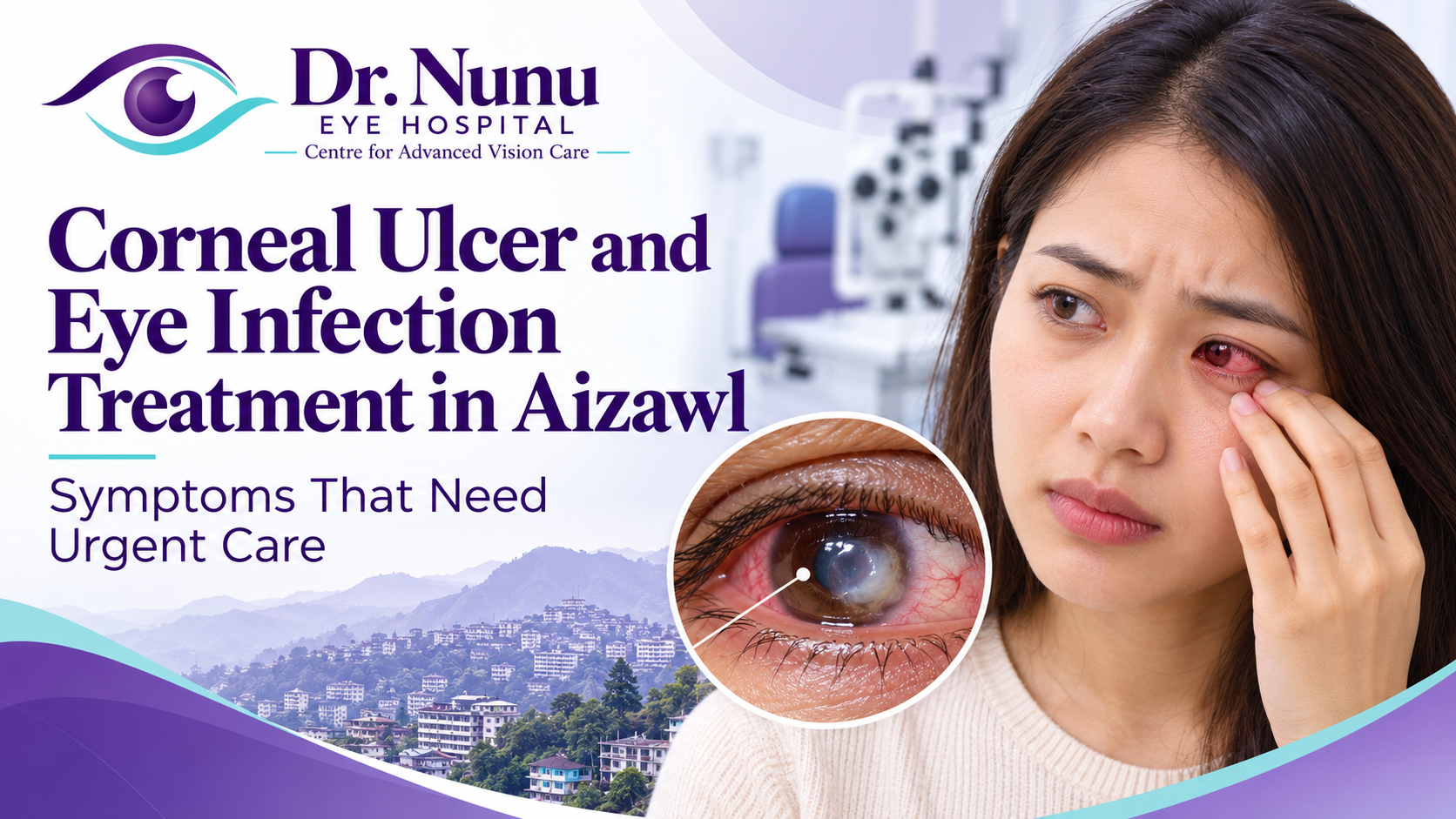
  • Corneal Ulcer and Eye Infection Treatment in Aizawl: Symptoms That Need Urgent Care

    Corneal Ulcer and Eye Infection Treatment in Aizawl: Symptoms That Need Urgent Care

    A painful red eye can be more than simple conjunctivitis. When the clear front surface of the eye—the cornea—becomes infected, inflammation and tissue damage can progress quickly and threaten vision. People seeking corneal ulcer and eye infection treatment in Aizawl should know which symptoms require a same-day eye examination and why using leftover drops can be dangerous.

    A corneal ulcer is an open sore or area of tissue breakdown in the cornea. Infectious keratitis means infection or inflammation of the cornea caused by bacteria, fungi, viruses or, less commonly, parasites. The terms overlap but are not identical. Both require professional assessment because the correct treatment depends on the cause, depth, location and severity.

    This guide explains urgent warning signs, risk factors relevant to contact-lens wearers and people with eye injuries, tests an ophthalmologist may perform, and the principles of treatment and follow-up. It is educational and cannot diagnose a red eye remotely.

    Why a corneal ulcer is an eye emergency

    The cornea is normally transparent and has an important role in focusing light. Infection can produce an inflammatory white or grey spot, an epithelial defect and swelling. Without timely treatment, an ulcer may deepen, scar the visual axis, thin or perforate the cornea, or allow infection to spread inside the eye.

    The National Eye Institute advises urgent help for intense eye pain, a change in vision, blurred vision, very red watery eyes, a retained object or serious trauma. These symptoms should not wait for a routine spectacle appointment.

    Symptoms that need urgent eye care

    Seek same-day professional assessment for any combination of:

    • moderate or severe eye pain;
    • rapidly increasing redness;
    • new blurred or reduced vision;
    • marked sensitivity to light;
    • continuous watering or thick discharge;
    • a foreign-body sensation that persists after rinsing;
    • a white, grey or cloudy spot on the cornea;
    • swollen eyelids with a painful eye;
    • difficulty opening the eye;
    • symptoms in a contact-lens wearer, especially after sleeping in lenses;
    • symptoms after plant matter, soil, dust, metal or chemical exposure;
    • pain that continues or worsens after contact-lens removal.

    Go to emergency care immediately for sudden major vision loss, penetrating injury, an object embedded in the eye, chemical burns, a visibly misshapen eye, severe trauma, or pain with nausea and coloured halos. For chemical exposure, begin continuous rinsing with clean water immediately and seek urgent help. Do not delay irrigation to search for a particular solution.

    Corneal ulcer versus conjunctivitis

    Conjunctivitis affects the thin membrane over the white of the eye and inner eyelids. It often causes redness, irritation and discharge. A corneal ulcer affects the clear corneal tissue and is more likely to cause significant pain, light sensitivity, reduced vision or a focal white spot.

    Symptoms can overlap, and it is unsafe to diagnose the cause from redness alone. Contact-lens wearers with a red or painful eye deserve particular caution because they have a higher risk of microbial keratitis. The CDC notes that contact-lens wearers with bacterial conjunctivitis are at increased risk of bacterial keratitis and need prompt ophthalmic evaluation.

    Common causes of corneal infection

    Bacterial keratitis

    Bacterial infection may progress rapidly. Contact-lens misuse is an important risk, but infection can also follow trauma, corneal surface disease, eyelid problems or reduced immunity. Treatment generally requires prescription antibiotic drops selected according to clinical findings and, in some cases, laboratory results.

    Fungal keratitis

    Fungal infection may follow injury with plant material, agricultural debris or soil, and can also occur with contact lenses or a compromised ocular surface. It may develop more gradually than some bacterial infections but can still cause severe damage. Ordinary antibiotic drops do not treat fungi.

    Viral keratitis

    Herpes simplex and herpes zoster viruses can involve the cornea. Episodes may recur. Treatment differs from bacterial or fungal disease, and steroid drops used without specialist direction can worsen some viral corneal infections.

    Acanthamoeba keratitis

    Acanthamoeba is a rare but serious infection associated particularly with contact lenses and water exposure. The CDC lists pain, redness, blurred vision, light sensitivity, foreign-body sensation and excessive tearing among possible symptoms. Showering, swimming or using a hot tub while wearing lenses can increase risk.

    Who is at higher risk?

    Risk factors include:

    • sleeping or napping in contact lenses not specifically prescribed for overnight wear;
    • wearing lenses longer than recommended;
    • using tap water on lenses or storage cases;
    • swimming or showering in contact lenses;
    • topping up old solution instead of using fresh disinfecting solution;
    • poor hand hygiene or a dirty lens case;
    • sharing cosmetic or decorative lenses;
    • eye injury, especially with soil or plant matter;
    • severe dry eye, eyelid disease or an unhealthy corneal surface;
    • previous corneal surgery or reduced corneal sensation;
    • diabetes, immune suppression or long-term steroid use;
    • unsupervised use of steroid-containing eye drops.

    In Mizoram, outdoor work, gardening and injury involving vegetation or soil are important details to mention during the consultation because they can influence the suspected organism and choice of tests.

    What contact-lens wearers should do immediately

    If a contact-lens wearer develops unusual redness, pain, light sensitivity, discharge or sudden blur, remove the lenses promptly and contact an eye doctor. Do not put the lens back in. The CDC describes microbial keratitis as a serious infection that can cause severe vision loss in the worst cases.

    Keep the lenses, case and solution rather than cleaning or discarding them before medical advice; the clinician may sometimes want this information. Use glasses until the eye doctor confirms it is safe to resume lens wear. Do not share drops or lenses.

    Tests for corneal ulcer and eye infection in Aizawl

    Accurate corneal ulcer and eye infection treatment in Aizawl begins with a careful diagnosis.

    A focused history and slit-lamp examination are central to diagnosis. The doctor asks when symptoms began, how quickly they changed, whether one or both eyes are affected, contact-lens habits, trauma, water exposure, previous surgery, medication and medical conditions.

    Vision assessment

    Each eye is tested separately. A reduction in vision helps indicate severity and provides a baseline for follow-up. Bring current spectacles if possible.

    Slit-lamp examination

    The slit lamp magnifies the cornea and front of the eye. The doctor assesses the size, depth and location of an infiltrate or ulcer, the surrounding swelling, inflammation inside the eye and any thinning. Central ulcers are especially concerning because scarring can directly affect vision.

    Fluorescein staining

    A temporary orange dye highlights breaks in the corneal surface under blue light. It helps measure the epithelial defect and identify scratches or characteristic staining patterns. The dye itself does not identify the organism.

    Corneal scraping and laboratory tests

    Some ulcers require a small sample from the corneal surface for microscopy and culture. Sampling is more likely when an ulcer is large, central, deep, atypical, associated with plant or water exposure, or not responding as expected. Results can guide a change in antimicrobial treatment.

    A scraping is performed using anaesthetic drops. Starting treatment may still be urgent, so the ophthalmologist balances timely medication with the need for useful samples.

    Additional imaging

    Photography can document size and healing. Anterior-segment OCT or ultrasound may be useful in selected cases, particularly when the cornea is very cloudy or complications are suspected. Not every patient needs imaging.

    Corneal ulcer and eye infection treatment in Aizawl

    A plan for corneal ulcer and eye infection treatment in Aizawl depends on the suspected cause and severity. Treatment should begin promptly when infection is suspected and may be adjusted after review or laboratory results.

    Prescription antimicrobial eye drops

    Bacterial disease is treated with antibiotic drops; fungal infection needs antifungal therapy; viral or Acanthamoeba disease requires different medicines. Severe infections may need drops very frequently, including during the night at the beginning. Follow the exact timetable and do not stop early because pain improves.

    Why steroid drops require caution

    Steroid drops can reduce inflammation in selected circumstances, but they can worsen or mask certain bacterial, fungal, viral and parasitic infections. Never begin a steroid or combination “red-eye” drop without an ophthalmologist’s instruction. If a clinician later adds a steroid, use it only at the prescribed stage and dose.

    Pain control and supportive care

    Oral pain relief may be advised according to the patient’s medical history. Lubrication or medicines that relax the focusing muscle may sometimes improve comfort. Patching the eye is not automatically appropriate for infection and should not be done unless specifically directed.

    Hospital care and surgery

    Admission may be considered for severe infection, rapid progression, inability to administer frequent drops, concern about perforation or difficulty attending close follow-up. Tissue adhesive, an amniotic membrane, corneal graft or other surgery may be required when there is advanced thinning, perforation, uncontrolled infection or significant structural damage.

    Why follow-up appointments are essential

    A corneal ulcer can change quickly. Early reviews may be daily or even more frequent depending on severity. The doctor checks pain, vision, defect size, infiltrate, thinning and inflammation. Treatment may be intensified, reduced or changed.

    Bring every prescribed bottle to follow-up so the team can confirm the correct medicine and technique. If symptoms worsen between visits, return earlier rather than waiting for the scheduled appointment.

    What not to do with a painful red eye

    • Do not use leftover antibiotic, steroid or combination drops.
    • Do not buy an anaesthetic eye drop for home use; repeated use can damage the cornea.
    • Do not wear contact lenses until cleared.
    • Do not rinse lenses or the eye with homemade salt water.
    • Do not place herbal products, honey, breast milk or other unsterile substances in the eye.
    • Do not cover a potentially infected eye tightly.
    • Do not delay because symptoms temporarily improve.

    Traditional or unprescribed remedies can contaminate the surface, delay effective treatment and make laboratory diagnosis more difficult.

    Preventing contact-lens-related infections

    Wash hands with soap and water and dry them before touching lenses. Rub and rinse reusable lenses with the recommended disinfecting solution, use fresh solution every time and never top off the case. Keep water away from lenses and cases. Remove lenses before swimming, showering or using a hot tub.

    Replace lenses and the case according to instructions. Do not sleep in lenses unless specifically prescribed, and understand that overnight wear still carries risk. Decorative lenses also require a professional prescription and fitting. See our guide to contact-lens fitting and safety in Aizawl.

    Preventing injury-related corneal infection

    Wear protective eyewear during gardening, construction, metalwork, carpentry, cutting vegetation and other jobs that send particles toward the face. After a minor loose particle enters the eye, rinse with clean water and avoid rubbing. If pain, blur or foreign-body sensation persists, seek care.

    Do not try to remove embedded metal, glass or plant material yourself. Penetrating injury requires emergency assessment. Tetanus protection and other treatment may be considered according to the type of injury.

    Can a corneal ulcer permanently affect vision?

    Yes. Healing can leave a scar, and the visual effect depends on its density and position. Central scarring may cause blur, glare or irregular astigmatism. Severe infection can lead to thinning, perforation, infection inside the eye or the need for transplantation. Early treatment improves the chance of controlling infection before extensive damage develops, but outcomes vary.

    After infection has resolved, later options may include spectacles, contact lenses designed for irregular corneas, laser treatment or corneal surgery, depending on the scar and eye health. These decisions are separate from the urgent phase.

    Frequently asked questions

    Is every red eye a corneal ulcer?

    No. Allergy, dryness, conjunctivitis, inflammation and glaucoma can also cause redness. Pain, light sensitivity, reduced vision, contact-lens wear or a visible corneal spot increase urgency, but examination is needed to identify the cause.

    Can I wait until morning?

    Severe pain, reduced vision, marked light sensitivity, a white corneal spot, trauma, chemical exposure or symptoms in a contact-lens wearer warrant urgent advice. If you are unsure, contact an eye-care service and describe the symptoms rather than delaying automatically.

    Can a pharmacist’s antibiotic drop treat it?

    The organism and severity cannot be reliably identified from symptoms alone. Some infections require specialised or very frequent medication, cultures and close review. Steroid-containing combination drops can be harmful in the wrong condition.

    Is a corneal ulcer contagious?

    The ulcer itself is not always passed from person to person, but some underlying viral or bacterial causes can spread. Wash hands, avoid sharing towels or cosmetics and follow the clinician’s instructions.

    When can I wear contact lenses again?

    Only after the infection has healed and the eye doctor approves. A new lens, case and solution may be recommended. The fit and care routine may need review before restarting.

    Will I need a corneal transplant?

    Most patients do not automatically need transplantation. Surgery is considered when infection cannot be controlled, the cornea becomes dangerously thin or perforates, or a healed scar causes severe visual impairment.

    Seek urgent eye infection care in Aizawl

    For corneal ulcer and eye infection treatment in Aizawl, early assessment is safer than watching a painful red eye at home. Dr. Nunu Eye Hospital & Research Centre can examine the cornea, assess urgency and advise on tests, prescription treatment and follow-up.

    Call +91 98620 19216 and clearly mention pain, reduced vision, light sensitivity, contact-lens wear, injury or a visible white spot so the clinic can advise on urgency. For penetrating trauma, chemical burns or sudden severe vision loss, seek emergency medical care immediately. You can also review our broader guide to emergency eye symptoms in Aizawl.

    Medical note: This article is general education, not a diagnosis or prescription. Corneal infection can threaten sight. Do not use antibiotics, steroids, anaesthetic drops or traditional remedies without professional examination.

  • Squint and Lazy Eye Treatment in Aizawl: Signs, Tests and Treatment for Children

    Squint and Lazy Eye Treatment in Aizawl: Signs, Tests and Treatment for Children

    A child’s eyes and vision develop rapidly during the early years of life. If one eye turns inward, outward, upward or downward, or if one eye does not develop clear vision, early assessment can make an important difference. Parents looking for squint and lazy eye treatment in Aizawl may hear the terms squint, strabismus and amblyopia used together, but they do not mean exactly the same thing.

    A squint, medically called strabismus, means the eyes are not aligned in the same direction. Amblyopia, often called lazy eye, means vision has not developed normally in one eye and occasionally in both eyes. A squint can cause amblyopia, but amblyopia may also develop because of unequal spectacle power, a cataract, a drooping eyelid or another condition that blocks clear visual input.

    This guide explains the signs families in Aizawl and across Mizoram should watch for, the tests commonly used for children, and treatments such as spectacles, patching, prescribed eye drops and squint surgery. Every child is different, so the treatment plan must follow an individual eye examination.

    What is a squint in children?

    A squint occurs when the two eyes do not point at the same target together. One eye may turn toward the nose, called an inward or convergent squint; drift toward the ear, called an outward or divergent squint; or move vertically. The turn may be constant, appear only at certain times, or alternate between the eyes.

    Some squints are obvious in photographs and face-to-face interaction. Others appear only when a child is tired, unwell, daydreaming or focusing at a particular distance. An intermittent squint still deserves assessment because its frequency, control and effect on binocular vision can change over time.

    Babies can occasionally appear cross-eyed during the first few months as visual coordination develops. However, a constant turn, an eye that does not move normally, or an intermittent turn that continues after about three months should be professionally assessed. The NHS guidance on squint similarly advises seeking help for a constant squint or one that comes and goes after three months of age.

    What is amblyopia or lazy eye?

    Amblyopia is reduced vision caused by abnormal visual development during childhood. The eye may look completely normal from the outside. The problem develops when the brain receives a clearer image from one eye than the other and increasingly relies on the stronger eye.

    According to the National Eye Institute, parents often do not know a child has amblyopia until it is found during an eye examination. Treatment is generally more effective in children than adults, which is why screening and early diagnosis matter.

    The word “lazy” can be misleading: the child is not choosing not to use the eye, and the eye muscles are not simply weak. Amblyopia is a problem in the way vision develops between the eye and brain. It should never be blamed on effort, behaviour or excessive screen use.

    How are squint and lazy eye connected?

    When a young child’s eyes point in different directions, the brain may suppress the image from the turned eye to avoid confusion or double vision. If suppression continues during the visual-development years, the sight in that eye may not reach its potential. This is strabismic amblyopia.

    However, not every child with a squint has amblyopia, and not every child with amblyopia has a visible squint. A large difference in spectacle prescription between the eyes can blur one image without changing eye alignment. Both eyes may also be affected if there is a high uncorrected prescription in each eye.

    Treatment has two related but distinct goals: improve vision in the weaker eye and improve or manage eye alignment. Patching can improve amblyopic vision, but it does not by itself permanently straighten a squint. Squint surgery changes eye alignment, but it does not automatically restore vision that failed to develop. Parents should understand which goal each recommended treatment addresses.

    Signs of squint and lazy eye in children

    Young children rarely describe blur in one eye because the stronger eye compensates. Families, teachers and caregivers may notice:

    • one eye turning inward, outward, upward or downward;
    • an eye turn that appears when the child is tired, ill or looking far away;
    • closing or covering one eye in bright light;
    • turning or tilting the head to look at objects;
    • sitting very close to a television or holding books near the face;
    • squinting, blinking or rubbing the eyes frequently;
    • difficulty following a moving object;
    • poor hand–eye coordination, catching difficulty or frequent bumping;
    • headaches, reading avoidance or loss of place while reading;
    • different reflections in the pupils in photographs;
    • an unusual white pupil reflex in a photograph.

    A head turn does not prove there is a squint, and sitting close to a screen does not by itself diagnose an eye condition. These clues indicate that an examination is worthwhile.

    Warning signs that need prompt assessment

    Arrange urgent medical assessment if a child develops a sudden new squint, especially with headache, vomiting, weakness, drooping eyelid, unequal pupils, unusual sleepiness, recent injury or restricted eye movement. A white or abnormal pupil reflex, an eye that appears enlarged, severe redness or pain, marked light sensitivity, or sudden loss of vision also needs prompt attention.

    A photograph showing a white pupil should not be dismissed as a camera effect until a clinician has assessed the child. Rare but serious eye conditions can present with an abnormal reflex or squint. If the problem follows trauma or chemical exposure, seek urgent care rather than waiting for a routine appointment. See our guide to emergency eye care in Aizawl.

    Why early assessment is important

    Visual development is most adaptable in childhood. Earlier identification gives clinicians more opportunity to provide a clear image to each eye and encourage the brain to use the weaker eye. This does not mean treatment should be abandoned in an older child; it means parents should avoid unnecessary delay and obtain an individual assessment.

    Untreated amblyopia can leave permanent reduced vision in the affected eye. A persistent squint may affect binocular vision and depth perception, and can have social or emotional effects. Timely care also helps identify spectacle needs or less common underlying problems.

    Tests used for squint and lazy eye treatment in Aizawl

    A paediatric eye assessment is adapted to the child’s age and ability. A child does not need to know letters or speak fluently for useful testing. Picture charts, matching tasks, lights, toys and objective instruments can provide valuable information.

    History and observation

    The clinician asks when the eye turn began, whether it is constant or intermittent, which eye turns and what makes it more noticeable. Premature birth, developmental history, illnesses, injury, previous glasses and family history of squint, amblyopia or childhood spectacles are relevant. Short videos or photographs taken at home can help when the turn is intermittent.

    Vision testing in each eye

    Each eye is assessed separately. Babies may be observed while following lights or objects. Toddlers can match pictures or symbols, while older children can use letters or numbers. Covering one eye at a time is essential because binocular testing may hide reduced vision in one eye.

    Cover tests and alignment measurement

    Covering and uncovering the eyes allows the examiner to observe refixation movements. Prisms may be used to measure the angle of the deviation at near and distance. Measurements may be repeated in different directions of gaze or while the child wears glasses.

    Eye-movement and binocular-vision tests

    The examiner checks whether both eyes move fully and together. Age-appropriate tests may assess fusion and stereopsis, or three-dimensional depth perception. These results help classify the squint and guide treatment.

    Cycloplegic refraction

    Special eye drops temporarily relax the focusing muscles and enlarge the pupils. This lets the clinician measure long-sightedness, short-sightedness and astigmatism more accurately, particularly in children who can focus strongly and hide part of their prescription. Near blur and light sensitivity can last for several hours after the drops.

    Eye-health examination

    The front and back of the eyes are examined to rule out cataract, retinal disease, optic-nerve problems or another cause of reduced vision. Dilation may be required. This health examination is an important reason not to rely only on a spectacle-shop screening or a photograph.

    Common causes and risk factors

    Sometimes no single cause is identified. Factors associated with childhood squint or amblyopia include:

    • uncorrected long-sightedness, short-sightedness or astigmatism;
    • a substantial difference in prescription between the eyes;
    • a family history of squint, amblyopia or early spectacles;
    • premature birth or low birth weight;
    • developmental or neurological conditions;
    • childhood cataract or drooping eyelid;
    • eye injury or disease that reduces clear visual input.

    Wide nasal bridges and skin folds at the inner eyelids can make a baby’s eyes appear crossed even when alignment is normal. This is sometimes called pseudo-squint. Because appearance alone cannot always distinguish a pseudo-squint from true strabismus, concerned parents should still arrange an examination.

    Squint and lazy eye treatment in Aizawl

    A plan for squint and lazy eye treatment in Aizawl should be personalised. The sequence and combination of treatment depend on the cause, age, vision in each eye, type of squint and how consistently it is present. Regular follow-up is important because prescriptions, alignment and visual development can change as a child grows.

    Spectacles

    Glasses create a clearer retinal image and are often the first treatment when a refractive error is present. In some long-sighted children, the correct glasses substantially reduce or fully control an inward eye turn. In amblyopia caused by unequal prescriptions, consistent spectacle wear alone may improve vision over time.

    Children may initially resist glasses. A well-fitting frame, positive routines and consistent wear usually help adaptation. Do not reduce wear or change the prescription without advice simply because the squint appears different with the glasses.

    Eye patching

    If vision remains weaker in one eye, the clinician may prescribe patching of the stronger eye for a specified number of hours. This encourages the brain to use the amblyopic eye. The dose varies; more is not automatically better and excessive unsupervised patching can be harmful.

    Patching should follow the exact schedule and be reviewed. Close-up activities such as drawing, puzzles or reading may keep a child engaged while patched if appropriate for their age. A patch treats reduced vision—it does not permanently straighten the eye turn.

    Atropine or other prescribed penalisation

    In selected cases, prescribed atropine drops can temporarily blur near vision in the stronger eye so the weaker eye is used more. The National Eye Institute notes that atropine may work as well as patching for some children. This is a medical treatment with potential side effects and must be used only under professional supervision.

    Eye exercises and prisms

    Exercises help only certain binocular-vision or eye-movement problems; they are not a universal treatment for childhood squint. Prisms may be useful in selected situations. Online exercises should not replace a diagnosis because different types of squint require different management.

    Squint surgery

    Surgery changes the position or tension of muscles on the outside of the eye to improve alignment. It may be considered when spectacles do not adequately control the turn or when the type and size of the squint make surgery appropriate. The eye is not removed from its socket.

    Surgery may improve alignment, binocular cooperation or an abnormal head position, but glasses can still be needed afterward. Amblyopia treatment may be required before or after surgery. Alignment outcomes are not perfectly predictable, and some children need more than one operation over time. The ophthalmologist should explain the expected benefit, anaesthesia, recovery, risks and alternatives for the individual child.

    Helping a child succeed with patching or glasses

    Young children often need time and encouragement. Explain treatment simply and positively. Let the child choose a patch design or spectacle frame, use a routine at the same time each day, and praise cooperation. Teachers and caregivers should know the schedule and understand that the child may initially find tasks harder while the stronger eye is covered.

    Keep a daily record of patch hours or drop use. If the child repeatedly removes the patch, develops a rash, cannot manage normal activities, or the family misses many doses, contact the clinic rather than changing the plan independently. Alternatives or practical adjustments may be possible.

    Preparing for a child’s eye appointment

    Bring current glasses, previous prescriptions, medicines and relevant medical reports. Note when the squint first appeared and whether it changes with fatigue, illness or viewing distance. Bring clear photographs or short videos showing the eye turn. Mention premature birth, developmental concerns and family eye history.

    Allow extra time if dilating drops are planned. Sunglasses or a hat may help with temporary light sensitivity afterward. Reassure the child that many tests use pictures, lights and games and that there are no wrong answers.

    Frequently asked questions

    Will my child grow out of a squint?

    Do not assume that a persistent or recurring squint will disappear without assessment. Some babies have a temporary appearance of crossing, but true squint can affect visual development and needs an eye examination.

    Is squint the same as lazy eye?

    No. Squint describes eye misalignment; lazy eye describes reduced visual development. They can occur together or separately.

    Can too much screen time cause a squint?

    Screen use may contribute to eye strain or reveal an existing intermittent problem, but it is not a complete explanation for a visible eye turn. A child with suspected squint needs examination rather than screen restriction alone.

    Does patching straighten the eyes?

    Patching is prescribed to improve vision in the weaker eye. It does not usually correct the underlying alignment permanently. Glasses, monitoring, exercises for selected conditions or surgery may address alignment.

    Will surgery cure lazy eye?

    Squint surgery repositions the eyes but does not directly restore amblyopic vision. The child may still need glasses, patching or prescribed drops.

    How long does treatment take?

    It varies from months to years depending on age, cause, severity and response. Regular appointments allow the clinician to adjust treatment and reduce it safely when vision stabilises.

    Should siblings be checked?

    A family history can increase risk. Ask an eye professional about appropriate screening, especially if a sibling has a squint, amblyopia or significant childhood prescription.

    Book a child eye assessment in Aizawl

    Families considering squint and lazy eye treatment in Aizawl should begin with a complete child eye assessment.

    If you notice an eye turn, abnormal head posture, unequal vision or another concern, arrange an examination rather than waiting for school screening. Dr. Nunu Eye Hospital & Research Centre can assess children in Aizawl and advise whether spectacles, amblyopia treatment, monitoring or further specialist management is appropriate.

    To enquire about squint and lazy eye treatment in Aizawl, call +91 98620 19216. Mention the child’s age, when the signs began and whether the change was sudden. Sudden squint with neurological symptoms, injury, severe pain or an abnormal white pupil requires urgent medical attention.

    Medical note: This article provides general education and does not diagnose an individual child. Treatment and timing depend on examination findings. Use patches, atropine or eye exercises only as prescribed and attend scheduled follow-up.

  • Comprehensive Eye Check-up in Aizawl: Tests, Cost Factors and When to Visit an Eye Doctor

    Comprehensive Eye Check-up in Aizawl: Tests, Cost Factors and When to Visit an Eye Doctor

    A comprehensive eye check-up in Aizawl does more than determine whether you need new spectacles. It evaluates how clearly each eye sees, how the eyes work together, the health of the cornea and lens, eye pressure, the optic nerve and, when appropriate, the retina. This matters because several eye diseases can develop quietly before a person notices significant vision loss.

    Whether you have blurred vision, headaches, diabetes, a family history of glaucoma or simply want to protect your sight, understanding the examination can make the visit easier. This guide explains common eye tests, factors that influence the cost of an examination and when adults and children in Aizawl and across Mizoram should consult an eye doctor.

    What is a comprehensive eye examination?

    A comprehensive eye examination is a structured assessment tailored to the patient’s age, symptoms, medical history and risk factors. It differs from a brief vision screening. A screening may identify that someone cannot see an eye chart clearly, but it may not explain why or evaluate the internal health of the eyes.

    The examination usually begins with a detailed history and visual-acuity testing. It may then include refraction, pupil and eye-movement assessment, slit-lamp examination, eye-pressure measurement and examination of the optic nerve and retina. Additional tests are selected only when the findings or medical history make them useful.

    The National Eye Institute explains that a dilated examination can detect eye diseases before they cause obvious vision loss. It may include visual acuity, visual field, eye-muscle, pupil-response and eye-pressure tests, followed by dilation to examine structures inside the eye.

    Why an eye-chart test alone is not enough

    Reading small letters measures central visual acuity, but good chart vision does not guarantee that every part of the eye is healthy. A person may read clearly while developing early glaucoma, diabetic retinal changes or peripheral retinal disease. Conversely, blurred chart vision may result from spectacle power, cataract, corneal disease, retinal disease or another cause.

    A comprehensive assessment connects visual performance with physical findings. It helps the doctor decide whether the patient needs spectacles, contact lenses, monitoring, treatment, imaging or referral for a particular procedure.

    Who should book a comprehensive eye check-up in Aizawl?

    Anyone with new or persistent visual symptoms should arrange an examination. Regular preventive examinations are also valuable for people who feel well, particularly when they have a higher risk of eye disease.

    Consider booking an appointment if you:

    • have blurred or fluctuating vision;
    • struggle to see road signs, the classroom board or fine print;
    • experience frequent headaches or eye strain;
    • notice glare, halos or difficulty seeing at night;
    • have red, painful, itchy, dry or watery eyes;
    • see flashes, new floaters, shadows or distorted lines;
    • have diabetes, high blood pressure or another relevant medical condition;
    • have a family history of glaucoma or inherited eye disease;
    • use contact lenses;
    • take medicines that may affect the eyes;
    • have had eye surgery or an eye injury;
    • are concerned about a child’s vision, eye alignment or school performance.

    Symptoms that should not wait for a routine appointment

    Sudden vision loss, a curtain-like shadow, severe eye pain, chemical exposure, penetrating injury, a painful red eye in a contact-lens wearer or flashes with a sudden increase in floaters may require urgent assessment. Do not wait for a planned routine check-up if symptoms are severe or rapidly changing.

    For more detail, read our guide to emergency eye care in Aizawl. After chemical exposure, begin rinsing the eye immediately with clean water and seek urgent professional care.

    What to bring to your eye appointment

    Bring your current spectacles, contact-lens packaging or prescription, and any eye drops or medicines you use. A list of medical conditions and family eye history is useful. Tell the clinic about previous eye surgery, injury, allergies, diabetes, blood-pressure problems and medication changes.

    If dilation may be needed, bring sunglasses because the eyes can be light-sensitive afterward. Near vision may remain blurry for several hours. Depending on your response to the drops and the type of work or driving you do, arranging transport or travelling with another person may be sensible.

    Tests during a comprehensive eye check-up in Aizawl

    Medical and eye-history review

    The doctor asks when symptoms began, whether they affect one or both eyes and whether they are constant or intermittent. Work, screen use, reading distance, driving, contact-lens habits and previous prescriptions provide context. General health is relevant because diabetes, hypertension, autoimmune disease and some medicines can affect the eyes.

    Distance and near visual acuity

    Each eye is tested separately using letters, numbers, symbols or age-appropriate pictures. Distance acuity shows how clearly you see far away, while near testing evaluates reading vision. Existing spectacles may be checked to measure how well they are working.

    Refraction and spectacle-power testing

    Refraction determines the lens power that provides the clearest vision. The examiner may use an automated reading as a starting point and then compare different lenses while asking which looks clearer. The final prescription also considers comfort, age, binocular balance, occupation and adaptation—not only a machine result.

    Children and some adults may need cycloplegic drops, which temporarily relax focusing. This can reveal hidden farsightedness or give a more reliable measurement when strong accommodation affects the result.

    Pupil examination

    The size, shape and reaction of the pupils provide information about the retina, optic nerve and neurological pathways. The doctor compares both eyes and checks their response to light. An abnormal response does not provide a diagnosis by itself, but it may guide further evaluation.

    Eye alignment and movement

    Eye-movement and cover tests assess whether the eyes point and move together. These are important for people with double vision, squint, reading difficulty or eye strain, and for children whose visual system is still developing.

    Slit-lamp examination

    A slit lamp is a microscope with a focused beam of light. It allows the doctor to examine the eyelids, lashes, conjunctiva, cornea, front chamber, iris and lens. It may reveal allergy, infection, dry-eye signs, corneal scars, inflammation and cataract.

    Fluorescein dye may be used to highlight damage to the corneal surface or assess tear-film stability. The dye is temporary and helps identify scratches, dry areas and patterns of surface disease.

    Eye-pressure measurement

    Tonometry measures pressure inside the eye. Different instruments use a gentle air puff, a small probe or a device that lightly contacts the numbed cornea. Eye pressure is an important part of glaucoma assessment, but a pressure reading alone cannot confirm or exclude glaucoma.

    The National Eye Institute notes that glaucoma detection requires a broader evaluation including history, visual acuity, pressure, optic-nerve and retinal examination, and visual-field testing when indicated. Some people develop glaucoma at statistically normal pressures, while many people with higher pressure never develop optic-nerve damage.

    Dilated retina and optic-nerve examination

    Dilating drops widen the pupils so the doctor can see more of the lens, vitreous, retina, retinal blood vessels and optic nerve. This examination can help identify diabetic retinopathy, retinal tears, macular disease, optic-nerve damage and other internal eye problems.

    Dilation is not necessarily required at every visit for every person. The doctor decides based on age, symptoms, risk factors and previous findings. When dilation is performed, temporary light sensitivity and blurred near vision are expected.

    Visual-field testing

    A visual-field test measures central and peripheral vision. A simple confrontation test may be performed during a routine examination. Automated field testing creates a more detailed map and may be recommended for suspected or known glaucoma, neurological symptoms or other conditions affecting side vision.

    Colour vision, depth perception and contrast

    These tests are selected when relevant. Colour testing may be useful for inherited colour-vision differences, optic-nerve concerns or occupational requirements. Depth-perception and binocular tests can help evaluate how the two eyes work together.

    When are OCT, corneal topography or retinal photographs needed?

    Not every patient needs imaging. Optical coherence tomography, commonly called OCT, uses light to create cross-sectional images of the retina or optic nerve. It may help evaluate glaucoma, macular disorders, diabetic changes and other retinal conditions.

    Retinal photographs document the appearance of the optic nerve and retina for review and comparison. Corneal topography maps the shape of the cornea and may be useful for irregular astigmatism, keratoconus, specialised contact lenses or surgical planning. Pachymetry measures corneal thickness and can contribute to glaucoma or corneal assessment.

    These tests complement the clinical examination; they do not replace the doctor’s interpretation. The reason for a test, what it can show and whether it is needed should be explained before it is performed.

    Cost factors for a comprehensive eye check-up in Aizawl

    The cost of a comprehensive eye check-up in Aizawl can vary because an examination is tailored to the patient. A straightforward vision assessment is different from a consultation that includes dilation, retinal imaging, visual fields or specialised corneal tests. Rather than relying on a generic price found online, ask the clinic what is included in the consultation and whether additional tests may carry separate charges.

    Type and purpose of the visit

    A routine spectacle check, medical eye consultation, contact-lens fitting, diabetic retinal review and pre-surgical evaluation involve different examination steps. Emergency assessments may also require immediate tests or treatment.

    Doctor consultation and clinical examination

    The base consultation may include history, acuity, refraction, slit-lamp examination and pressure measurement, but inclusions differ between facilities. Ask whether refraction, dilation and a written prescription are included.

    Additional diagnostic testing

    OCT, visual fields, corneal topography, retinal photography, ultrasound or other tests can affect the total cost. These should be recommended according to clinical need rather than added automatically to every examination.

    Dilation and follow-up

    Some clinics include dilation or a review visit in the consultation; others charge separately depending on the service. Certain findings may require a follow-up after additional testing or after beginning treatment.

    Glasses, contact lenses and medicines

    The consultation fee is separate from the cost of spectacle frames, lenses, contact lenses, solutions or prescribed medicines unless the clinic specifically states otherwise. Lens material, coatings, prescription strength and frame choice can create a wide range of optical costs.

    Insurance and documentation

    Coverage depends on the patient’s policy, provider network and whether the service is considered routine, diagnostic or surgical. Confirm requirements directly with the insurer and clinic. Bring identification, referral documents and previous reports when applicable.

    How often should adults have an eye examination?

    There is no single interval appropriate for everyone. The timing depends on age, symptoms, spectacle or contact-lens use, family history, medical conditions and previous findings. A healthy young adult with no risk factors may need a different schedule from an older adult with diabetes or a person being monitored for glaucoma.

    The National Eye Institute advises people to discuss their personal risk with an eye doctor. Those with diabetes, hypertension or other risk factors may need regular dilated examinations, often at least annually depending on medical guidance. Patients with established eye disease may need more frequent visits.

    Do not wait for the next planned interval if vision changes. New symptoms should determine the urgency of review.

    Eye examinations for people with diabetes

    Diabetes can damage retinal blood vessels before vision becomes noticeably blurred. The Centers for Disease Control and Prevention recommends regular dilated examinations so diabetic retinal disease can be identified early. The appropriate timing depends on the type and duration of diabetes, pregnancy, retinal findings and the treating clinicians’ advice.

    Good blood-glucose, blood-pressure and cholesterol management supports eye health but does not replace retinal examination. Read more about diabetic retinopathy screening in Aizawl.

    Eye check-ups for children

    Children may not realise that their vision is blurred because they have no clear vision to compare it with. Parents and teachers should watch for squinting, sitting close to screens, holding books near the face, closing one eye, an eye turning, frequent headaches, rubbing, loss of place while reading or declining classroom performance.

    Children with premature birth, developmental concerns, a family history of childhood eye disease or visible eye abnormalities may need earlier and more frequent assessment. School screening is helpful but does not replace a complete examination when symptoms or risk factors are present.

    Our guide to childhood myopia in Aizawl explains common signs of short-sightedness and why monitoring progression matters.

    Eye check-ups after age 40

    Near focusing commonly becomes more difficult in the forties because of presbyopia. New reading difficulty may be corrected with spectacles, but an examination also provides an opportunity to review eye pressure, the optic nerve, lens clarity and retinal health.

    Risk for cataract, glaucoma, diabetic eye disease and macular problems generally increases with age. Gradual change can be easy to overlook when both eyes compensate for each other, so testing each eye separately is important.

    What happens after the examination?

    The doctor should explain the findings and next steps. Possible outcomes include reassurance, a spectacle prescription, advice about screen habits or dry-eye care, medication, additional testing, monitoring, contact-lens assessment or discussion of a procedure.

    If spectacles are prescribed, ask whether they are intended for distance, reading, computers, full-time use or a specific task. If a disease is identified, understand the follow-up interval, warning symptoms and treatment goal. Keep copies of prescriptions and reports so changes can be compared over time.

    Frequently asked questions about eye check-ups in Aizawl

    Will every comprehensive eye check-up include dilation?

    No. Dilation depends on age, symptoms, medical history, risk and what the undilated examination shows. It is particularly valuable when the retina, optic nerve or internal eye structures need detailed assessment.

    Can I drive after my pupils are dilated?

    Dilation can cause glare, light sensitivity and blurred near vision for several hours. Effects vary. Ask the clinic in advance and arrange transport if you may not be comfortable or safe driving afterward.

    Should I stop wearing contact lenses before the appointment?

    Instructions depend on the reason for the visit and lens type. A corneal measurement or new fitting may require lenses to be removed for a specified period. Ask the clinic when booking and bring the lenses, case, solution and packaging.

    Does normal eye pressure mean I do not have glaucoma?

    No. Pressure is only one part of glaucoma assessment. Optic-nerve appearance, visual fields, corneal thickness, drainage angle, family history and change over time may also matter.

    Do I need an eye examination if my spectacles still feel fine?

    Possibly. Spectacles correct focus but do not screen for every eye disease. Examination timing should reflect age, medical risks, family history and previous findings, even when vision seems stable.

    Why can the final cost differ between patients?

    Patients may need different tests. One person may require only a consultation and refraction, while another needs dilation, OCT, visual fields or corneal mapping. Request an estimate based on the planned examination and ask before optional or additional tests are performed.

    Book a comprehensive eye check-up in Aizawl

    Dr. Lynapawngia Solo at Dr. Nunu Eye Hospital & Research Centre provides eye assessments based on symptoms, age, visual needs and medical risk. The objective is not simply to issue a spectacle prescription, but to understand how well the eyes see and whether the visible and internal structures appear healthy.

    To enquire about a comprehensive eye check-up in Aizawl, call +91 98620 19216 during available clinic hours. Bring current spectacles, medicines and previous reports. Mention sudden or severe symptoms when booking so the clinic can advise on urgency.

    Medical note: This article provides general education and does not replace an individual eye examination. Tests, fees and follow-up requirements vary according to clinical need. Seek urgent care for sudden vision loss, severe pain, chemical injury, penetrating trauma or rapidly worsening symptoms.

  • Eye Allergy Treatment in Aizawl, Mizoram: Causes of Itchy, Red and Watery Eyes

    Eye Allergy Treatment in Aizawl, Mizoram: Causes of Itchy, Red and Watery Eyes

    People seeking eye allergy treatment in Aizawl often report itchy, red and watery eyes, but the cause is not always the same. In many people, these symptoms are caused by eye allergy, also called allergic conjunctivitis. Others may have dry eye, eyelid inflammation, an infection, irritation from smoke or cosmetics, or a problem related to contact lenses. Because treatments differ, a careful eye examination is important when symptoms are severe, recurrent or unclear.

    This guide explains eye allergy treatment in Aizawl, common triggers in Mizoram, how doctors distinguish allergy from other red-eye conditions, and practical steps that may reduce symptoms safely.

    What is an eye allergy?

    The conjunctiva is the thin, transparent tissue covering the white part of the eye and the inner surface of the eyelids. Allergic conjunctivitis develops when this tissue reacts to a substance that the immune system treats as a threat. The reaction releases inflammatory chemicals, including histamine, which can cause itching, redness, swelling and tearing.

    Eye allergy often affects both eyes and may occur with sneezing, a runny or blocked nose, asthma, eczema or other allergic conditions. It is not contagious. However, viral and bacterial conjunctivitis can also make eyes red and watery, so it is unwise to assume that every red eye is an allergy.

    The Centers for Disease Control and Prevention notes that viruses, bacteria and allergens are among the common causes of conjunctivitis. Chemical irritation, foreign bodies and contact-lens wear can produce similar symptoms.

    Typical symptoms of allergic conjunctivitis

    Itching is the symptom most strongly associated with eye allergy. People may describe an intense urge to rub, burning, grittiness or a feeling of tired eyes. Other possible symptoms include:

    • redness of the whites of the eyes;
    • clear, watery tearing;
    • puffy or swollen eyelids;
    • mild stringy mucus;
    • sensitivity to bright light;
    • symptoms in both eyes;
    • sneezing, nasal congestion or an itchy nose.

    Symptoms may appear soon after exposure to a trigger or build gradually. Rubbing often brings brief relief but can worsen inflammation, swelling and surface irritation. Persistent rubbing is especially concerning in children and young people because repeated mechanical stress is associated with corneal problems.

    Common triggers considered in eye allergy treatment in Aizawl

    Aizawl’s green hills, changing seasons, indoor dampness, road dust, smoke and household exposures can all affect sensitive eyes. Individual triggers vary, and the presence of a substance does not prove that it is the cause. A symptom diary can help identify patterns.

    Pollen and plant material

    Airborne pollen from grasses, weeds and trees may trigger seasonal symptoms. Windy days, gardening, outdoor work and travel through vegetation can increase exposure. Pollen counts and flowering periods vary by plant and weather, so symptoms may not follow the same schedule every year.

    House dust and dust mites

    Dust accumulates in bedding, mattresses, upholstered furniture, curtains, books and soft toys. Dust mites thrive in humid indoor environments and may cause symptoms throughout the year. Sweeping or shaking bedding can temporarily send particles into the air.

    Mould and dampness

    Mould spores may be present in damp rooms, bathrooms, storage areas, walls with water damage and poorly ventilated spaces. Mizoram’s humid and rainy periods can make moisture control challenging. Visible mould or a musty smell should prompt attention to leaks, ventilation and safe cleaning.

    Animal dander

    Proteins from skin flakes, saliva and urine of cats, dogs and other animals may trigger eye and nasal symptoms. The allergen can remain on clothing, bedding and furniture even when the animal is not in the room.

    Smoke, dust and air irritants

    Vehicle exhaust, construction dust, wood smoke, tobacco smoke, incense, aerosol sprays and strong fragrances may irritate eyes directly or worsen an existing allergy. Irritation is not always a true immune allergy, but the symptoms can look similar.

    Cosmetics, skincare and eye medicines

    Makeup, lash products, hair dye, facial creams, cleansers and preservatives in some eye drops can irritate the eye or cause contact allergy around the eyelids. A new product is an important clue, but reactions can also develop after repeated use of a familiar product.

    Types of eye allergy

    Seasonal allergic conjunctivitis

    This common form appears during periods when particular pollens or outdoor allergens are present. Symptoms may begin suddenly and occur with hay-fever symptoms.

    Perennial allergic conjunctivitis

    Perennial allergy may persist throughout the year, often linked with dust mites, mould, animals or other indoor triggers. Symptoms can be milder but more continuous.

    Vernal keratoconjunctivitis

    Vernal keratoconjunctivitis is a more serious recurrent allergic condition seen especially in children and young people. It can produce intense itching, thick mucus, marked light sensitivity and changes beneath the upper eyelid. The cornea may be involved, so specialist assessment and ongoing treatment are important.

    Atopic keratoconjunctivitis

    This chronic condition is often associated with atopic dermatitis or eczema. It may affect the eyelids, conjunctiva and cornea and can threaten vision if severe or poorly controlled.

    Giant papillary conjunctivitis

    This is commonly associated with mechanical irritation from contact lenses, deposits on lenses or occasionally exposed sutures. Wearers may notice itching, mucus, reduced lens tolerance or excessive lens movement. Lens wear should be stopped and the eyes assessed.

    Eye allergy or infection: how can you tell?

    Allergy commonly causes strong itching, clear tearing and involvement of both eyes. Viral conjunctivitis may begin in one eye and spread to the other, often with watery discharge, a recent respiratory illness or contact with someone who has red eyes. Bacterial conjunctivitis is more likely to produce pus-like discharge and eyelids that stick together, although symptoms overlap.

    Eye pain, significant light sensitivity or reduced vision is not typical of simple allergy and needs prompt assessment. A contact-lens wearer with a painful red eye should remove the lenses and seek urgent eye care because corneal infection can progress rapidly.

    Do not start leftover antibiotic drops. Antibiotics do not treat allergy or most viral conjunctivitis, and inappropriate drops can cause toxicity, allergy or delay the correct diagnosis.

    Other conditions that can resemble eye allergy

    Dry eye can cause burning, fluctuating vision, watering and redness. Paradoxically, an unstable tear film can produce excessive reflex tears. Blepharitis and meibomian-gland dysfunction may cause crusting, sore eyelids and irritation. A foreign body, corneal scratch, uveitis, glaucoma and other eye diseases can also present with redness.

    Watery eyes may result from a blocked tear-drainage pathway. Eyelid dermatitis can be triggered by cosmetics or skin products. Because these problems require different treatment, recurrent “allergy” that does not improve should be re-evaluated rather than repeatedly self-treated.

    How an eye doctor evaluates itchy, red and watery eyes

    Assessment begins with the timing of symptoms, whether one or both eyes are affected, known allergies, contact-lens use, medicines, occupation, home environment and recent exposure to infection. The doctor asks about pain, discharge, light sensitivity and visual change.

    Visual acuity is checked, and a slit-lamp microscope is used to examine the eyelids, conjunctiva, cornea and tear film. The pattern of redness, papillae beneath the eyelids, mucus, corneal staining and lid-margin changes can help identify the cause. Severe, unusual or persistent cases may need further testing or coordination with an allergy or medical specialist.

    Eye allergy treatment in Aizawl

    Treatment depends on the diagnosis, severity, age, other health conditions and whether the cornea is involved. Mild symptoms may improve with trigger reduction, cold compresses and preservative-free lubricating drops. Moderate or recurrent allergy may require specific anti-allergy medication prescribed or recommended after examination.

    Avoid or reduce the trigger

    Complete avoidance is not always possible, but reducing exposure can lower the allergic load. Close windows during dusty or high-pollen periods when practical, wash the face after outdoor exposure, and change clothes after gardening or dusty work. Wear well-fitting glasses outdoors to reduce direct exposure.

    For indoor triggers, wash bedding regularly, reduce dust-collecting clutter, ventilate damp areas and repair leaks. Clean visible mould safely and address the source of moisture. Keep animals out of the bedroom if pet dander is a suspected trigger.

    Cold compresses

    A clean, cool compress placed gently over closed eyelids can ease itching and swelling. Use a separate clean cloth for each person. Do not apply ice directly to the skin or eye.

    Lubricating eye drops

    Artificial tears may dilute allergens and soothe the ocular surface. Preservative-free products may be preferable when drops are used frequently or when the surface is sensitive. Not every “redness relief” product is a lubricant, so ask an eye-care professional which type is suitable.

    Anti-allergy eye drops

    Antihistamine and mast-cell-stabilising drops target the allergic response. Some products combine both actions. Choice and frequency depend on the individual, and children, pregnant patients, people with glaucoma and those using other eye medicines should seek professional advice.

    The CDC’s guidance on conjunctivitis treatment notes that removing the allergen and certain topical or systemic allergy medicines can relieve allergic conjunctivitis. A clinician can help select appropriate treatment.

    Oral allergy medicines

    Oral antihistamines may help when eye symptoms occur with nasal allergy, but some can worsen dryness. They should be used according to medical or pharmacy advice, particularly in children or people taking other medication.

    Steroid eye drops require supervision

    Steroid drops can be valuable for selected severe allergic eye disease, but they must not be self-started. Unsupervised use may raise eye pressure, contribute to cataract, worsen herpes infection or mask other disease. Treatment requires an eye examination and monitoring.

    What not to do when your eyes itch

    • Do not rub the eyes, even though rubbing may feel temporarily satisfying.
    • Do not share eye drops, towels, pillows, makeup or contact lenses.
    • Do not put rose water, honey, breast milk, saliva, herbal preparations or non-sterile liquids into the eyes.
    • Do not use an old prescription simply because symptoms seem similar.
    • Do not use steroid-containing drops without an eye doctor’s supervision.
    • Do not wear contact lenses while the eyes are red, painful or producing discharge.

    The National Eye Institute describes cold compresses and appropriate over-the-counter drops as possible supportive measures for mild conjunctivitis, but the correct approach depends on the cause. Home care should never delay assessment when warning signs are present.

    Contact lenses and eye allergy

    Allergens and protein deposits can collect on contact lenses, while inflamed eyes may tolerate lenses poorly. Remove lenses during an active episode and use spectacles. Do not resume wear until symptoms have settled and an eye-care professional says it is safe, especially if pain, discharge or corneal involvement occurred.

    Follow the prescribed replacement schedule, use fresh recommended solution and never expose lenses or cases to water. Daily disposable lenses may reduce deposit and care issues for some patients, but a professional contact lens fitting in Aizawl is needed to decide whether they are suitable.

    Eye allergies in children

    Children may rub, blink or squeeze their eyes instead of describing itching. Parents may notice redness, watering, swollen lids, dark circles or stringy mucus. Ask about sneezing, eczema, asthma and seasonal patterns.

    Persistent rubbing, strong light sensitivity, difficulty opening the eyes, a white spot on the cornea or reduced vision requires prompt examination. Severe childhood allergic eye disease can affect the cornea and should not be managed only with over-the-counter drops.

    Prevention after eye allergy treatment in Aizawl

    Wash hands before touching the face. Keep frequently used bedding and towels clean, and avoid sharing them. Clean spectacles regularly. When sweeping, construction work or smoke exposure cannot be avoided, protective eyewear may reduce direct irritation.

    During long screen sessions, blink fully and take regular breaks because dry-eye symptoms can add to allergic discomfort. Keep fans and vehicle vents from blowing directly into the eyes. Store eye drops as directed and avoid touching the bottle tip to the eyelids or lashes.

    A diary noting location, weather, activity, cosmetics, animals and symptom severity can reveal patterns. Bring the record and all eye medicines to the appointment.

    When to seek urgent eye care

    Arrange prompt or same-day assessment if redness is accompanied by:

    • eye pain or severe discomfort;
    • marked sensitivity to light;
    • blurred or reduced vision;
    • a white or cloudy spot on the cornea;
    • injury, chemical exposure or a suspected foreign body;
    • thick discharge or rapidly increasing swelling;
    • symptoms in a contact-lens wearer;
    • headache, nausea or coloured halos;
    • symptoms that worsen or fail to improve;
    • redness in a newborn baby.

    The CDC advises medical review for conjunctivitis with pain, light sensitivity, persistent blurred vision, intense redness or worsening symptoms. For serious warning signs, see our guide to emergency eye care in Aizawl.

    Frequently asked questions about eye allergy treatment in Aizawl

    Is eye allergy contagious?

    No. Allergic conjunctivitis itself does not spread between people. Viral and bacterial conjunctivitis can be contagious, which is why diagnosis matters when family members, classmates or colleagues develop similar symptoms.

    Can an eye allergy affect only one eye?

    Allergy commonly affects both eyes, but symptoms may be unequal. Persistent one-sided redness should be examined to exclude a foreign body, infection, tear-drainage problem or another cause.

    Why do my eyes water if allergy also makes them feel dry?

    Surface irritation can trigger reflex tearing, while the quality and stability of the tear film remain poor. Allergy and dry eye can occur together and may need a combined treatment plan.

    Are redness-removing drops safe every day?

    Some vasoconstrictor drops temporarily shrink surface blood vessels without treating the underlying cause, and redness can rebound with repeated use. Ask an eye-care professional before regular use.

    Will antibiotics treat itchy eyes?

    Antibiotics treat specific bacterial infections; they do not treat allergic conjunctivitis. Unnecessary antibiotics may irritate the surface or cause an allergic reaction.

    Can eye allergy damage vision?

    Simple seasonal allergy usually does not damage vision, but severe forms such as vernal or atopic keratoconjunctivitis can involve the cornea. Pain, light sensitivity or reduced vision requires prompt assessment.

    Book an eye-allergy assessment in Aizawl

    Dr. Lynapawngia Solo at Dr. Nunu Eye Hospital & Research Centre can examine itchy, red and watery eyes, identify features that suggest allergy or another cause, and recommend treatment based on the findings.

    To enquire about eye allergy treatment in Aizawl, call +91 98620 19216 during available clinic hours. Bring any eye drops you currently use, your spectacles or contact-lens details, and a list of possible triggers.

    Medical note: This article provides general education and does not replace an individual eye examination. Seek prompt professional care for pain, light sensitivity, reduced vision, injury, chemical exposure, severe swelling or rapidly worsening redness.

  • Contact Lens Fitting in Aizawl: Types, Eye Tests, Safety and Care

    Contact Lens Fitting in Aizawl: Types, Eye Tests, Safety and Care

    Contact lenses can provide clear vision without spectacles, but they are not one-size-fits-all products. A lens rests directly on the tear film and must match the shape, health and visual needs of each eye. A professional contact lens fitting in Aizawl therefore involves more than checking spectacle power or choosing a lens from a packet.

    During a fitting, the eye-care professional examines the front surface of the eyes, measures the cornea, reviews the tear film, discusses work and lifestyle needs, selects a trial lens and checks how it behaves on the eye. Follow-up is important because a lens that feels comfortable for a few minutes may not remain healthy or stable throughout the day.

    This guide explains the main types of contact lenses, the eye tests involved, safe wearing habits and warning signs that patients in Aizawl and across Mizoram should know.

    Why professional contact lens fitting matters

    A spectacle prescription is not the same as a contact lens prescription. Contact lenses sit much closer to the eye, and their effective power may differ, especially with stronger prescriptions. A contact lens specification may also include the lens material or brand, base curve, diameter, replacement schedule and other design details.

    Two lenses with the same printed power can differ in shape, thickness, oxygen transmission, edge design and surface properties. These differences can affect comfort, movement, vision and corneal health. Buying a lens based only on spectacle power or changing brands without professional advice can lead to blur, dryness, poor fit or complications.

    International safety guidance treats contact lenses, including decorative lenses, as medical devices. The US Food and Drug Administration advises people to use a current prescription and receive regular eye checks. Local prescribing rules may differ, but the clinical principle is relevant everywhere: lenses should be selected and monitored for the individual eye.

    Who may benefit from contact lenses?

    Contact lenses can correct common refractive errors such as myopia, hyperopia, astigmatism and presbyopia. They may be helpful for sports, social occasions, certain occupations, unequal prescriptions between the eyes or patients who prefer a wider field of view than spectacles provide.

    Some people require specialised lenses for irregular corneas, keratoconus, significant astigmatism, corneal scars, dry-eye conditions or after certain eye procedures. Contact lenses can also have therapeutic uses under specialist supervision.

    Not everyone is an immediate candidate. Active infection, significant uncontrolled allergy, severe dryness, poor eyelid hygiene, inability to handle lenses safely or difficulty following the care schedule may need attention first. The decision depends on examination findings rather than age or preference alone.

    Types considered during contact lens fitting in Aizawl

    Soft contact lenses

    Soft lenses are flexible and usually adapt quickly to the eye. They are available in different materials, including hydrogel and silicone hydrogel. Silicone hydrogel designs generally allow more oxygen to pass through the lens, but material choice still depends on tear film, comfort, prescription and wearing pattern.

    Soft lenses may be designed for daily disposable use or planned replacement over a longer period. Daily disposables are discarded after one day and do not require a storage case or overnight disinfection. Reusable lenses must be cleaned and disinfected exactly as directed and replaced on schedule.

    Toric lenses for astigmatism

    Toric contact lenses contain different powers in different meridians and use stabilisation features to maintain the correct orientation. The fitter assesses rotation and stability after the lens settles. A lens that rotates excessively may produce fluctuating blur even when its power appears correct.

    Multifocal and monovision lenses

    Presbyopia commonly affects near focusing from the forties onward. Multifocal contact lenses combine distance and near optics in one design. Monovision uses one eye mainly for distance and the other mainly for near tasks. Both strategies require careful trial and realistic expectations because adaptation, lighting and visual demands vary between patients.

    Rigid gas-permeable lenses

    Rigid gas-permeable, or RGP, lenses are smaller and retain their shape on the eye. They can provide excellent optics for some patients with high astigmatism or irregular corneas. Initial awareness may be greater than with soft lenses, so adaptation and correct handling are important.

    Hybrid and scleral lenses

    Hybrid lenses combine a rigid central zone with a soft outer skirt. Scleral lenses are larger rigid lenses that vault over the cornea and rest on the white part of the eye. They create a fluid reservoir and may be considered for selected irregular-cornea or ocular-surface conditions. These are specialised fittings that need detailed measurements, trial evaluation and follow-up.

    Coloured and decorative lenses

    Cosmetic lenses alter eye colour or appearance, but they still contact the cornea and require proper fitting. The FDA’s decorative-lens guidance warns that improperly fitted lenses can cause scratches, infection and reduced vision. Never share decorative lenses, and avoid sellers who do not require appropriate eye assessment or prescription information.

    Eye tests before prescribing contact lenses

    Medical and lifestyle history

    The fitting begins with questions about previous lens wear, allergies, dryness, eye surgery, medicines and general health. Diabetes, autoimmune disease and some medications may influence the ocular surface. The clinician also asks about screen time, travel, sports, dust exposure, working hours and how many days or hours the lenses will be worn.

    Visual acuity and refraction

    Visual acuity measures how clearly each eye sees. Refraction identifies the lens power needed to correct myopia, hyperopia and astigmatism. For strong prescriptions, the contact lens power may be adjusted because the lens sits directly on the eye rather than several millimetres in front of it.

    Slit-lamp examination

    A slit lamp provides a magnified view of the eyelids, lashes, conjunctiva, cornea and tear film. The clinician looks for inflammation, allergy, dryness, scars, abnormal blood vessels and other findings that could affect safe wear. Baseline examination also makes later changes easier to identify.

    Corneal measurements

    Keratometry measures corneal curvature. Corneal topography may create a more detailed map, especially when the cornea is irregular or a specialised lens is being considered. These measurements help guide the starting lens design, but a successful fit must still be observed directly on the eye.

    Tear-film assessment

    The tear film supports comfort, lubrication and optical quality. The professional may evaluate tear quantity, tear stability, blinking and the oil glands along the eyelid margins. Dry-eye or eyelid problems may need treatment before or alongside contact lens wear.

    Pupil, iris and eyelid assessment

    Pupil size can influence glare, halos and multifocal performance. Iris diameter, eyelid position, blink pattern and lid tension may affect lens selection or movement. These details are particularly relevant for specialised, toric or multifocal designs.

    What happens during the trial-lens assessment?

    After selecting an initial lens, the clinician places it on the eye and allows time for settling. Vision is checked, and additional power may be tested over the trial lens. The lens is viewed with the slit lamp to assess centration, coverage, movement with blinking and response to eye movements.

    A soft lens should normally cover the cornea, centre appropriately and move enough to support tear exchange without being excessively loose. A lens that is too tight may initially feel comfortable but move poorly. A loose lens may decentre, cause variable vision or irritate the eyelids. Rigid and scleral lenses are evaluated using criteria appropriate to their design.

    Toric lenses are marked so rotation can be measured. Multifocal lenses may require adjustments based on distance, computer and reading tasks. Specialised fittings sometimes require several trials, and this is a normal part of finding the safest and most useful option.

    Insertion, removal and handling training

    A proper contact lens fitting in Aizawl includes teaching the patient to insert and remove lenses without injuring the eye. The patient should demonstrate the technique independently before taking lenses home.

    Wash hands with soap and dry them completely using a clean, lint-free towel. Check that the lens is clean, intact and not inside out. Work over a clean surface and keep fingernails away from the eye. If a lens falls onto an unclean surface, follow the correct cleaning or disposal instructions for that lens type.

    Do not use saliva to wet a lens. Do not share lenses, cases or solution. If a lens is torn, chipped or persistently uncomfortable, remove it rather than trying to tolerate it.

    Safe schedules after contact lens fitting in Aizawl

    Build wearing time according to the schedule given by the eye-care professional. Do not exceed the recommended hours or replacement interval simply because a lens still feels comfortable. A daily disposable lens is used once; it is not designed for cleaning and reuse. Monthly or other reusable lenses should be replaced according to the prescribed interval after opening, not only after a certain number of wears.

    Sleeping in lenses increases risk unless a particular lens and schedule have been specifically prescribed for overnight use. Even approved extended wear carries additional risk and requires supervision. When in doubt, remove lenses before sleeping or napping.

    Cleaning and storing reusable lenses

    Use only the care system recommended for your lens type. The Centers for Disease Control and Prevention advises rubbing and rinsing reusable lenses with appropriate fresh solution and avoiding “topping off” old solution in the case.

    Empty the case after each use, rub and rinse it with fresh disinfecting solution, and allow it to air-dry upside down with the caps off on a clean tissue. Replace the case regularly according to professional and manufacturer advice. Saline can rinse lenses in specific routines but does not disinfect them.

    Hydrogen-peroxide systems require the special neutralising case supplied with the product and the complete stated neutralisation time. Unneutralised peroxide should never be placed directly in the eye.

    Keep contact lenses away from water

    Remove lenses before showering, swimming or entering a hot tub. Do not rinse lenses or cases with tap, filtered, bottled or well water. Water can make soft lenses swell or adhere to the eye and can introduce organisms that cause serious corneal infection.

    The CDC’s contact-lens water safety advice notes the risk of Acanthamoeba, a microorganism found in many water sources. If lenses are exposed to water, remove them as soon as possible and follow professional or manufacturer guidance about discarding or properly disinfecting them.

    Contact lenses, screens and Aizawl’s environment

    Long screen sessions reduce blink frequency and may make lenses feel dry. Follow regular screen breaks, blink fully, maintain suitable working distance and use only lubricating drops approved for contact-lens wear. Airflow from fans, heaters or vehicle vents directed at the face may also increase dryness.

    Dust, smoke, seasonal allergy and travel can affect comfort. Carry spectacles and a clean lens case when appropriate. Do not keep wearing a painful lens because removing it is inconvenient. Patients travelling within Mizoram should pack adequate prescribed supplies and avoid transferring solution into unlabelled containers.

    Warning signs: remove the lenses and seek advice

    Remove contact lenses immediately if you develop increasing redness, pain, light sensitivity, discharge, sudden blur, unusual watering or the sensation that something remains in the eye. Do not put the lens back in to see whether symptoms settle.

    Contact-lens wear is a recognised risk factor for microbial keratitis, an infection of the cornea that can progress rapidly. Same-day examination is important when pain, redness and reduced vision occur together. Severe pain, rapidly worsening symptoms or major vision loss may require emergency assessment. See our guide to emergency eye care in Aizawl.

    Avoid using leftover antibiotic, steroid or redness-relief drops without examination. Steroids can worsen some infections, and symptom-masking drops do not address the cause.

    Follow-up after contact lens fitting in Aizawl

    Follow-up allows the clinician to check vision, comfort, wearing time, lens condition and the health of the cornea and conjunctiva. Attend even when the lenses feel comfortable, because early changes are not always obvious to the wearer.

    Wear the lenses for the requested period before the visit unless they cause pain or other warning symptoms. Bring the lens packaging, case, solution and spectacles. Explain honestly how long the lenses are worn, whether naps occur and how cleaning is performed; accurate information helps the clinician make safer recommendations.

    A change in prescription, brand, material, medication, pregnancy, work environment or eye health may require reassessment. Never assume that an old contact lens specification remains appropriate indefinitely.

    Common questions about contact lens fitting in Aizawl

    Can I use my spectacle power to buy contact lenses?

    No. Spectacle power is only one part of the decision. Contact lens power, material, base curve, diameter and design must be assessed on the eye.

    Are daily disposable lenses always best?

    They reduce care steps and case-related contamination, but suitability, power availability, cost and ocular findings vary. The best lens is the one that meets visual needs while remaining healthy and practical for the patient.

    Can I wear lenses if I have dry eyes?

    Some patients with dryness can wear selected lenses after tear-film and eyelid assessment, treatment and schedule adjustment. Others may need to delay or limit wear. Self-selecting a “high-water” lens does not guarantee comfort.

    Can teenagers wear contact lenses?

    Age alone does not determine suitability. Hygiene, motivation, handling ability, parental support and eye health matter. Children and teenagers should be supervised and should always have backup spectacles.

    Can coloured lenses be worn without vision power?

    Even plano decorative lenses require correct measurement and health assessment because their shape and material still affect the eye.

    Book a contact lens assessment in Aizawl

    Dr. Lynapawngia Solo at Dr. Nunu Eye Hospital & Research Centre can assess vision, corneal health, tear film and suitability for contact lenses. A structured fitting helps match the lens to the eye and provides the training needed for safer daily use.

    To enquire about contact lens fitting in Aizawl, call +91 98620 19216 during available clinic hours. Bring your current spectacles, previous contact lens details and any eye medicines to the appointment.

    Medical note: This article provides general education and does not replace an individual eye examination. Lens availability and suitability vary. Remove lenses and seek prompt professional care for pain, marked redness, light sensitivity, discharge or reduced vision.

  • Emergency Eye Care in Aizawl: Symptoms That Need Immediate Treatment

    Emergency Eye Care in Aizawl: Symptoms That Need Immediate Treatment

    Eye problems can change quickly. A person may feel comfortable in the morning and develop pain, redness or loss of vision later in the day. Some symptoms can safely wait for a scheduled eye examination, but others may signal a condition in which early treatment is important for protecting sight. Knowing when to seek emergency eye care in Aizawl can help patients and families make a safer decision.

    Sudden vision loss, a chemical splash, a penetrating injury, severe pain, a curtain-like shadow, or a painful red eye should never be ignored. These symptoms do not identify one specific disease, and it is not possible to judge their seriousness accurately at home. This guide explains common warning signs, basic first aid, what not to do, and what patients in Aizawl and across Mizoram can expect during an urgent eye examination.

    If you currently have sudden loss of vision, a serious injury, chemical exposure, severe pain, or eye symptoms with weakness, facial drooping, confusion or difficulty speaking, seek the nearest appropriate emergency medical service immediately.

    What is an eye emergency?

    An eye emergency is a symptom or injury that may threaten vision, damage the eye, or point to a serious medical or neurological condition. The eye is delicate, and problems affecting the cornea, retina, optic nerve, eye pressure or blood supply may require assessment much sooner than a routine spectacle appointment.

    Not every red or watery eye is an emergency. Mild irritation without pain or vision change may have a less urgent cause. However, pain, reduced vision, light sensitivity, trauma, recent surgery or contact-lens use can change the level of concern. When you are uncertain, it is safer to obtain professional advice than to delay because the eye “does not look too bad.”

    Symptoms that need immediate emergency eye care in Aizawl

    1. Sudden loss or major reduction of vision

    A sudden change in vision in one or both eyes is an emergency even when it is painless. A person may describe complete darkness, a missing area, heavy blur, distorted sight, faded colours or vision that disappears and then returns.

    Possible causes include a retinal problem, blockage of blood flow, optic-nerve disease, bleeding inside the eye, acute glaucoma or a neurological event. Do not wait for the vision to clear on its own, and do not drive yourself. Note the exact time the change began because timing may affect investigation and treatment.

    2. New flashes, many floaters or a curtain-like shadow

    Floaters may look like dots, threads, cobwebs or small shadows that move with the eyes. A few longstanding floaters can be harmless. A sudden shower of new floaters, flashes of light, peripheral blur or a dark curtain across vision can indicate a retinal tear or retinal detachment.

    The National Eye Institute advises immediate assessment when new floaters appear suddenly with flashes or a dark shadow because retinal tear or detachment can be a medical emergency. Risk can be higher in people with high myopia, diabetes, an eye injury or previous cataract surgery.

    3. Severe eye pain or a very painful red eye

    Severe pain, especially when accompanied by redness, blurred vision, halos, headache, nausea or vomiting, requires urgent evaluation. Causes may include acute angle-closure glaucoma, corneal infection, inflammation inside the eye, injury or another condition that cannot be distinguished by appearance alone.

    Do not use leftover antibiotic or steroid drops. Steroid drops can worsen certain infections and should be used only after an appropriate examination. If pain is strong enough to disturb sleep or is combined with loss of vision, go to an emergency medical facility.

    4. Chemical splash in the eye

    Household cleaners, lime, cement, laboratory chemicals, agricultural products and industrial substances can injure the ocular surface. Chemical exposure is a true emergency because damage may continue while the substance remains in contact with the eye.

    Begin rinsing immediately with plenty of clean, gently flowing water. Do not delay irrigation while searching for a special solution or calling a clinic. Hold the eyelids open, blink during rinsing and remove contact lenses if they come out easily. Continue rinsing while arranging emergency care. Do not attempt to neutralise an acid with an alkali or an alkali with an acid.

    Guidance from the American Academy of Ophthalmology’s EyeWiki emphasises that immediate, copious irrigation is the cornerstone of chemical-burn treatment and should not be delayed to identify the chemical. Bring the product container or a photograph of its label if this can be done safely.

    5. A cut, puncture or object embedded in the eye

    A high-speed fragment from hammering, grinding, cutting metal, construction work or an explosion can penetrate the eye even when the external wound appears small. Glass, wire, wood and sharp tools can also cause serious injury.

    Do not press, rub or rinse an eye if you suspect it has been cut or punctured. Do not attempt to pull out an embedded object. Avoid putting pressure from a cloth patch over the eye. Protect it loosely from further contact and proceed to an emergency department for urgent ophthalmic assessment.

    6. Blunt trauma to the eye or face

    A ball, fist, fall, road accident or other blunt impact can cause bleeding, fracture, retinal injury or damage to the lens even when the eyelids are not cut. Seek urgent help for reduced vision, double vision, severe pain, an irregular pupil, blood visible inside the eye, restricted eye movement or increasing swelling.

    A cold compress may be held gently over the surrounding area for a minor bruise, but do not apply pressure directly to the eyeball. A significant injury needs examination rather than home observation.

    7. Contact-lens wear with pain, redness or blurred vision

    Contact-lens wearers can develop corneal infection, which may progress quickly. Remove the lenses promptly if you experience unusual irritation, redness, increasing pain, light sensitivity, discharge or sudden blur. Do not put the lens back in and do not rely on redness-relief drops.

    The Centers for Disease Control and Prevention advises contact-lens users with these symptoms to remove their lenses and contact an eye doctor immediately. Bring the lenses, case and solutions to the appointment if requested, but do not delay care to locate them.

    8. Eye symptoms after surgery or an injection

    Some mild irritation may occur after an eye procedure, but severe or increasing pain, worsening redness, sudden blur, discharge, flashes, numerous floaters or rapidly decreasing vision can indicate a complication. Contact the operating team immediately or seek emergency assessment, particularly if the procedure was recent.

    Do not stop prescribed postoperative medicines unless instructed, but do not add other drops on your own. Take the discharge summary and medication list with you.

    9. Sudden double vision or eye symptoms with neurological signs

    New double vision can arise from an eye-muscle, nerve or neurological problem. It needs urgent evaluation when it starts suddenly or occurs with severe headache, a drooping eyelid, unequal pupils, weakness, numbness, facial drooping, confusion, imbalance or difficulty speaking.

    These combined symptoms may require general emergency or stroke assessment rather than only an eye-clinic visit. Note the time of onset and use emergency medical services; do not drive yourself.

    10. Eye emergencies in children

    Children may be unable to describe vision loss or the exact site of pain. Seek prompt help after a chemical splash, sharp-object injury, high-speed impact, animal scratch or suspected foreign body. Warning signs include refusal to open an eye, persistent crying, strong light sensitivity, an abnormal pupil, blood, marked swelling or suddenly bumping into objects.

    Do not force the eyelids open or allow the child to rub the eye. Keep medicines and cleaning products in their original containers and out of children’s reach.

    Eye symptoms that are urgent even without severe pain

    The seriousness of an eye condition is not always proportional to discomfort. Retinal detachment and some blood-vessel problems may initially be painless. Seek same-day advice for a new missing area in vision, sudden distortion, abrupt colour change, transient darkness, a new curtain or shadow, or a sudden increase in floaters.

    People with diabetes should be particularly alert to sudden blur or new floaters because bleeding or other retinal complications may occur. Routine screening remains important even when vision feels normal; see our guide to diabetic retinopathy screening in Aizawl and Mizoram.

    First aid before seeking emergency eye care in Aizawl

    First aid depends on the type of emergency. The following measures are general guidance and do not replace professional assessment:

    • Chemical exposure: start immediate, continuous rinsing with clean water and arrange emergency care.
    • Loose surface particle: blink and allow natural tears to help. Gentle rinsing may be reasonable if there is no high-speed injury or suspected penetration.
    • Embedded or penetrating object: do not remove, rinse, press or manipulate it. Protect the area from contact and seek emergency care.
    • Blunt injury: avoid pressure on the eyeball. A gentle cold compress around the area may help a minor bruise, but vision change or significant impact needs examination.
    • Contact-lens symptoms: remove the lenses, stop wearing them and seek prompt advice for pain, redness, light sensitivity or blur.
    • Sudden vision loss or neurological signs: record the onset time and use an emergency medical service.

    What not to do during an eye emergency

    • Do not rub or press the injured eye.
    • Do not attempt to remove an embedded object.
    • Do not delay chemical irrigation while searching for medicine.
    • Do not use homemade remedies, saliva, honey or unsterile liquids.
    • Do not use another person’s eye drops.
    • Do not self-start steroid drops.
    • Do not patch an eye tightly after a possible penetrating injury.
    • Do not wear contact lenses over a painful or red eye.
    • Do not drive when vision is suddenly reduced or double.
    • Do not eat or drink excessively when urgent surgery or sedation might be required.

    What to expect from emergency eye care in Aizawl

    The clinical team will ask when the symptoms began, whether one or both eyes are affected, and whether there was an injury, chemical, contact lens, operation or general-health problem. Provide the exact substance or mechanism whenever possible.

    Assessment may include visual-acuity testing, pupil reactions, eye movements, slit-lamp examination, fluorescein dye, eye-pressure measurement and a dilated retinal examination. Pressure testing may be avoided when an open-globe injury is suspected. Retinal imaging, visual-field assessment, ultrasound, CT scanning or blood tests may be required depending on the findings.

    The purpose is to identify the anatomical structure involved and decide whether the patient needs medicine, a procedure, surgery, imaging, observation or transfer to a facility with appropriate emergency resources.

    Emergency eye care versus a routine appointment

    Emergency eye care in Aizawl is intended for sudden or serious symptoms, while a routine appointment is generally suitable for gradual spectacle-power changes, longstanding mild dryness, stable eyelid symptoms or a planned screening examination. Urgent assessment is more appropriate when symptoms are sudden, severe, linked to an injury or operation, or accompanied by vision loss.

    A red eye with mild itching and no pain may result from allergy, but a red eye with pain, light sensitivity or reduced vision needs quicker evaluation. Our separate article explains common causes and treatment of conjunctivitis in Aizawl.

    How to prepare while travelling for urgent treatment

    If possible, ask a relative or friend to accompany you. Bring current medicines, allergy information, spectacles, previous reports and details of recent surgery. For chemical exposure, bring the container or a clear photograph of the label without delaying irrigation.

    Patients travelling from outside Aizawl should not postpone time-sensitive emergency care for a long journey when a nearer emergency department can provide initial assessment and stabilisation. The safest destination depends on the condition, time of day and available local services.

    Preventing serious eye injuries

    Many eye injuries are preventable. Wear appropriate protective goggles during grinding, welding, construction, carpentry, farming, laboratory work, chemical handling and activities that can create flying fragments. Ordinary spectacles do not provide the same protection as safety eyewear.

    Store cleaning chemicals safely, read labels and avoid transferring corrosive products into drink bottles. Supervise children around sharp objects, fireworks, elastic projectiles and household chemicals. Contact-lens users should wash and dry their hands, follow replacement schedules, avoid sleeping or swimming in lenses unless specifically approved, and never rinse lenses with tap water.

    Seeking emergency eye care in Aizawl

    Dr. Lynapawngia Solo at Dr. Nunu Eye Hospital & Research Centre assesses eye symptoms and helps patients determine the appropriate next step. For urgent advice during available clinic hours, call +91 98620 19216.

    For sudden blindness, a penetrating injury, chemical burn, severe trauma, stroke-like symptoms or another major emergency, proceed to the nearest equipped emergency department or use local emergency medical services without waiting for a routine clinic appointment.

    Medical note: This article provides general information and does not diagnose an individual condition. Emergency services, opening hours and available specialist care can change. Severe or sudden symptoms require immediate assessment at an appropriate medical facility.