
Clear vision. Compassionate care. Better tomorrow.Call: +91 98620 19216
☎ Call now

Author: Dr. Lynapawngia Solo

  • Eye Hospital in Aizawl: A Complete Guide to Eye Tests, Doctors and Treatment

    Eye Hospital in Aizawl: A Complete Guide to Eye Tests, Doctors and Treatment

    Finding the right eye hospital in Aizawl is important whether you need a routine vision test, treatment for an uncomfortable eye problem, management of a long-term condition, or an assessment for surgery. Eye symptoms can affect people of every age, and the appropriate care depends on the cause rather than the symptom alone.

    Blurred vision, redness, watering, headaches, eye pain and difficulty reading can have many possible explanations. Some problems are minor and temporary, while others require prompt examination to protect sight. Conditions including glaucoma, cataract and diabetic retinopathy may also progress before a person notices a major change in vision.

    This guide explains when to visit an eye doctor, what happens during an eye examination, which services patients may need and how families in Aizawl and other parts of Mizoram can prepare for an appointment.

    Why choosing an eye hospital matters

    An eye hospital does much more than check spectacle power. A comprehensive eye examination evaluates vision and looks for abnormalities involving the eyelids, tear film, cornea, lens, optic nerve and retina.

    Many eye diseases do not cause clear warning signs during their early stages. The National Eye Institute notes that an eye examination may be the only reliable way to confirm that the eyes are healthy when a condition has not yet produced noticeable symptoms.

    An ophthalmologist is a medically trained eye doctor who can diagnose eye diseases, prescribe treatment and perform eye surgery when required. Patients may also receive support from optometrists, technicians, nurses and counsellors as part of their care.

    Selecting an eye hospital in Aizawl with access to appropriate examination equipment and specialist assessment makes it easier to move from diagnosis to treatment without unnecessary delays.

    When should you visit an eye doctor in Aizawl?

    You do not have to wait until your vision becomes severely blurred. Schedule an eye examination when you experience:

    • Persistent or recurring blurred vision
    • Difficulty reading signs, books or phone screens
    • Frequent headaches during close work
    • Red, itchy, burning or watery eyes
    • Eye pain or discomfort
    • Sensitivity to light
    • Difficulty seeing at night
    • Halos around lights
    • A change in spectacle power
    • Flashes, floaters or shadows in vision
    • Double vision
    • An eye injury or chemical exposure
    • Vision problems associated with diabetes
    • A family history of glaucoma or another significant eye disease

    Regular examinations may also be advisable for children, older adults, people who use spectacles or contact lenses, patients with diabetes and anyone already receiving treatment for an eye condition.

    The frequency of follow-up should be personalised. Age, symptoms, general health, family history, eye pressure and previous examination findings all affect how often a patient should return.

    Symptoms that require urgent assessment

    Some symptoms should not be treated as a routine spectacle problem. Seek urgent medical care if you experience sudden loss or marked reduction of vision, a dark curtain or shadow moving across your vision, new flashes accompanied by numerous floaters, severe eye pain, a painful red eye, an eye injury, a chemical splash, sudden persistent double vision, or vision changes accompanied by weakness, confusion, facial drooping or difficulty speaking.

    The Centers for Disease Control and Prevention advises people not to wait for a routine appointment when they develop decreased vision, eye pain, double vision, flashes, floaters or halos around lights.

    If chemicals enter the eye, begin rinsing immediately with clean water and obtain emergency medical help. Do not rub an injured eye or attempt to remove an embedded object yourself.

    What happens during an eye checkup?

    The exact tests depend on your age, symptoms and medical history. A comprehensive assessment commonly begins with questions about the problem, when it started and whether it affects one or both eyes.

    Tell the doctor about diabetes, high blood pressure, thyroid disease, allergies, previous eye surgery, injuries and medicines. Bring your current spectacles, prescriptions and relevant medical reports.

    Vision testing

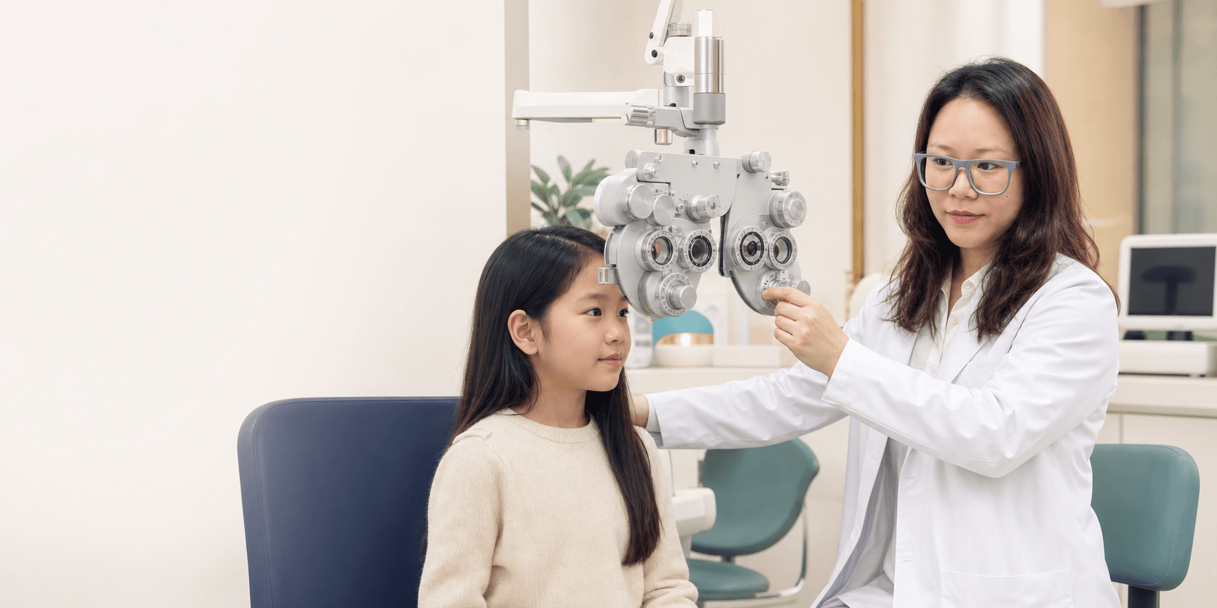
    Visual acuity testing measures how clearly each eye sees. Distance and near vision may be tested separately. Children and adults who cannot read standard letters can be examined using alternative symbols or matching tests.

    Testing each eye individually is important because one stronger eye can sometimes hide reduced vision in the other.

    Refraction and spectacle power

    Refraction determines whether myopia, hyperopia, astigmatism or presbyopia is affecting vision. The doctor or optometrist compares different lenses to find the power that provides the clearest and most comfortable result.

    A change in power may explain blurred vision or headaches, but clear vision with lenses does not rule out every eye disease. The health of the eye still needs to be assessed.

    Eye pressure measurement

    Eye pressure testing can form part of glaucoma assessment. A pressure reading is useful, but glaucoma cannot be diagnosed or excluded by pressure alone. Doctors may also examine the optic nerve, visual field, drainage angle and corneal thickness when clinically indicated.

    Slit-lamp examination

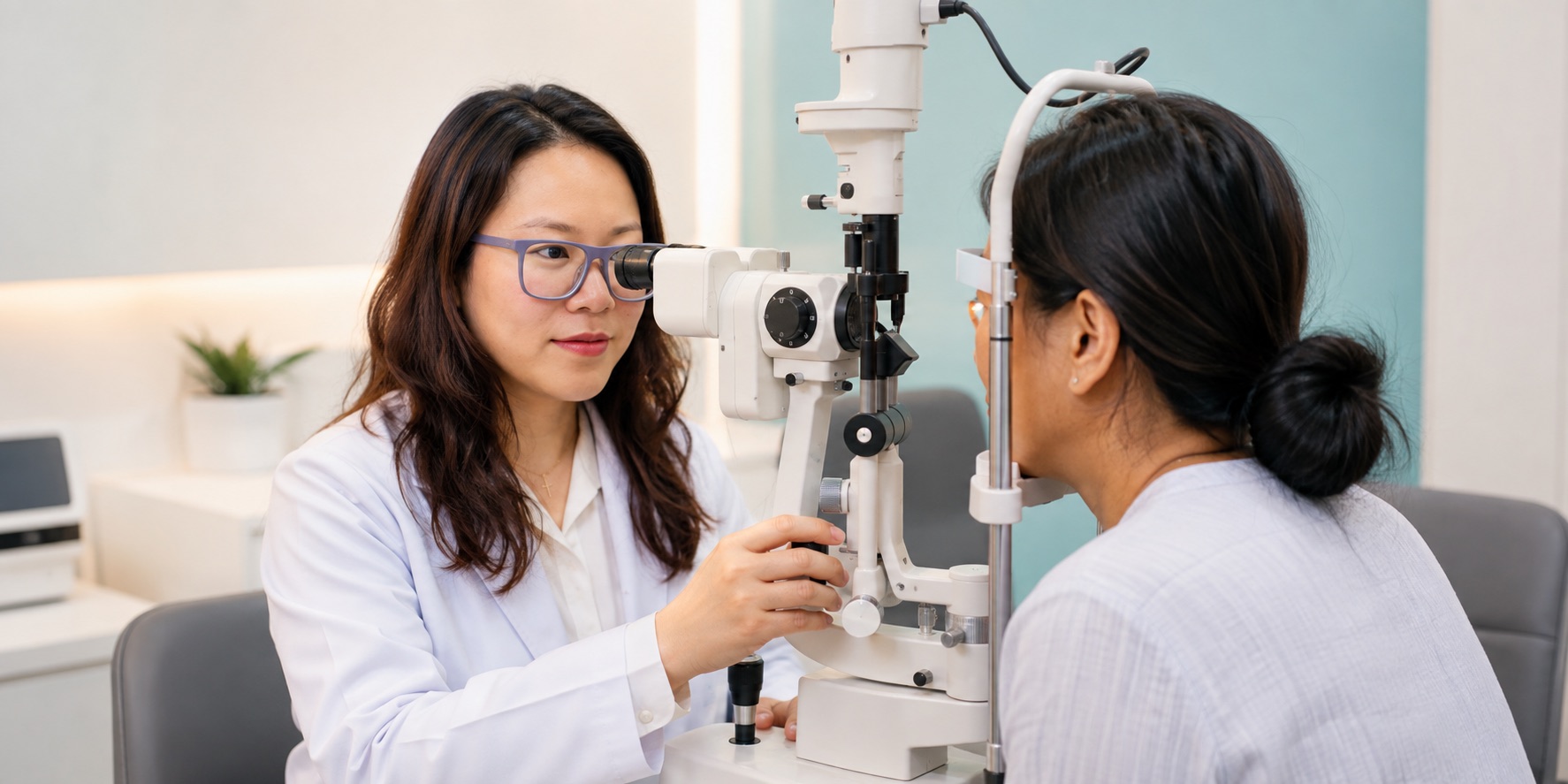
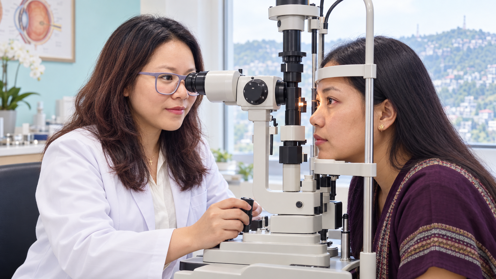
    A slit lamp is a microscope that allows the doctor to inspect the front structures of the eye in detail. It can help identify dry eye, eyelid inflammation, allergy, corneal problems, infection, injury and cataract.

    Dilated retinal examination

    Eye drops may be used to enlarge the pupils so the doctor can examine the lens, optic nerve, retina and retinal blood vessels. Dilation is particularly valuable when assessing diabetes-related changes, retinal symptoms and certain unexplained vision problems.

    Vision may remain blurred at near distance and sensitive to light for several hours afterward. Patients should ask whether they need someone to accompany them, especially if they have not been dilated before.

    Eye conditions commonly assessed at an eye hospital

    Cataract

    A cataract develops when the natural lens inside the eye becomes cloudy. Common symptoms include gradual blurred vision, glare, faded colours and difficulty driving or seeing in dim light.

    Early cataracts may be managed by updating spectacles and improving lighting. When the cataract meaningfully interferes with daily activities, the doctor may discuss surgery. The decision should be based on the patient’s functional difficulty, eye health and personal needs—not age alone. Read our detailed guide to cataract surgery in Aizawl.

    Glaucoma

    Glaucoma is a group of conditions that damage the optic nerve. Some forms progress gradually without obvious early symptoms, making examination especially important for people with risk factors.

    Treatment may include eye drops, laser procedures or surgery. Patients already using glaucoma medication should not stop it without consulting their doctor. Learn more about glaucoma screening in Aizawl and Mizoram.

    Diabetic retinopathy

    Diabetes can damage the small blood vessels of the retina. Early diabetic retinopathy may not noticeably affect sight, so retinal examinations remain important even when vision appears normal.

    Good management of blood glucose, blood pressure and cholesterol supports overall health but does not replace an eye examination. Depending on the findings, treatment can include observation, injections, laser procedures or surgery. Read our guide to diabetic retinopathy screening in Aizawl.

    Dry eye

    Dry eye can cause burning, grittiness, fluctuating blur, watering and tired eyes. Screen use, reduced blinking, air conditioning, contact lenses, eyelid problems and some medicines may contribute.

    Treatment depends on the underlying pattern and severity. It may involve environmental changes, eyelid care, lubricating drops or prescription treatment. Avoid repeatedly using steroid or redness-relief drops without medical supervision. See our patient guide to dry-eye treatment in Aizawl.

    Refractive errors

    Myopia, hyperopia, astigmatism and presbyopia affect the way light is focused. They can cause blurred vision, headaches, squinting or difficulty changing focus.

    Spectacles are a common and effective solution. Some patients may also be suitable for contact lenses or surgical vision-correction options following a complete assessment.

    Conjunctivitis and eye infections

    Redness, discharge, itching and watering can result from infection, allergy, dryness or irritation. Because these causes require different treatments, antibiotic drops should not be used automatically for every red eye.

    Pain, light sensitivity, contact-lens use or reduced vision can indicate a more serious problem and warrant prompt assessment. Read more about conjunctivitis treatment in Aizawl.

    Childhood eye problems

    Children may not realise that their vision is abnormal. Parents and teachers should watch for squinting, sitting close to screens, covering one eye, frequent eye rubbing, headaches or difficulty seeing the classroom board.

    Early examination is especially important for suspected amblyopia, commonly called lazy eye, because treatment is most effective during childhood. The CDC reports that amblyopia affects approximately two to three out of every 100 children.

    How to choose an eye hospital in Aizawl

    A practical choice should be based on the type and quality of care required rather than advertising alone. Consider whether the facility offers:

    • Evaluation by an eye-care professional
    • Comprehensive eye examinations and refraction
    • Slit-lamp examination
    • Eye pressure and glaucoma assessment
    • Retinal and diabetic eye assessment
    • Cataract evaluation and surgical counselling
    • Care for children and older adults
    • Clear explanations of findings and treatment choices
    • Suitable follow-up after treatment or surgery
    • Accessible appointment information

    A trustworthy facility should explain which tests are needed, what the results mean, the available treatment alternatives and when follow-up is necessary.

    Questions to ask an ophthalmologist

    1. What is causing my symptoms?
    2. Does the problem affect one or both eyes?
    3. Do I need spectacles, medicine, further tests or surgery?
    4. What may happen if the condition is not treated?
    5. Which warning signs require urgent help?
    6. How should I use the prescribed medicine?
    7. Could diabetes or another health condition be contributing?
    8. When should I return for follow-up?
    9. Should close relatives have their eyes examined?
    10. Are there temporary restrictions on driving, work or screen use?

    If surgery is being considered, ask about its purpose, benefits, alternatives, recovery period, possible complications and follow-up schedule.

    How to prepare for your appointment

    Bring your current spectacles, previous prescriptions, medical reports and a list of medicines. Tell the doctor if you have diabetes, high blood pressure, allergies, previous eye surgery or a family history of eye disease.

    Write down when your symptoms began, whether they are constant, and what makes them better or worse. If discomfort is connected with computer work, note the type of device, approximate viewing distance and daily screen hours.

    Dilating drops may temporarily blur near vision and increase light sensitivity. Ask in advance whether someone should accompany you.

    Protecting your eyes between examinations

    • Take regular breaks during prolonged screen work.
    • Wear appropriate eye protection during hazardous work.
    • Manage blood glucose and blood pressure with your physician.
    • Do not share eye drops or use someone else’s prescription.
    • Keep contact lenses and their cases clean.
    • Avoid repeated use of steroid drops without supervision.
    • Follow the prescribed schedule for glaucoma or other long-term treatment.
    • Seek help promptly after an eye injury or sudden vision change.

    Healthy habits support vision, but they cannot replace an examination when symptoms persist or a person has risk factors for eye disease.

    Eye care for patients across Mizoram

    Patients travelling to an eye hospital in Aizawl from another part of Mizoram may benefit from planning ahead. Confirm clinic hours, bring relevant records and ask whether dilation or diagnostic testing may be required.

    If an appointment is related to possible surgery, ask whether preliminary tests and follow-up visits can be coordinated. Patients with sudden vision loss, severe pain or major injury should seek the nearest emergency medical facility rather than delaying treatment for travel.

    Visit Dr. Nunu Eye Hospital & Research Centre

    Dr. Nunu Eye Hospital & Research Centre provides eye examinations and specialist ophthalmic care for patients in Aizawl and across Mizoram. Dr. Lynapawngia Solo assesses patients with blurred vision, eye discomfort, cataract, glaucoma concerns, diabetes-related risks, childhood vision problems and other eye-health needs.

    If you are looking for an eye hospital in Aizawl for a routine checkup or a specific concern, arrange an examination so the cause can be identified and the appropriate next step discussed.

    For appointments, call +91 98620 19216.

    Medical note: This article provides general educational information and does not replace an individual medical examination or diagnosis. Sudden vision loss, severe eye pain, injury or neurological symptoms require urgent medical attention.
  • Digital Eye Strain Treatment in Aizawl, Mizoram: Symptoms and Prevention

    Digital Eye Strain Treatment in Aizawl, Mizoram: Symptoms and Prevention

    Phones, laptops, tablets, televisions, and desktop computers are now part of study, work, communication, banking, entertainment, and everyday life. After long periods of screen use, many people notice tired eyes, headaches, burning, dryness, watering, or temporary blur. These symptoms are often described as digital eye strain or computer vision syndrome. For anyone searching for digital eye strain treatment in Aizawl, the first step is to understand why the symptoms occur and whether a simple change in screen habits is enough.

    Digital eye strain is usually uncomfortable rather than dangerous, but similar symptoms can also come from an incorrect spectacle prescription, dry-eye disease, allergy, an eye-muscle coordination problem, migraine, or another condition. Persistent, severe, or one-sided symptoms deserve an eye examination. This detailed guide from Dr. Lynapawngia Solo explains the causes, practical prevention, eye tests, and treatment options for adults, students, children, and families in Aizawl and across Mizoram.

    What is digital eye strain?

    Digital eye strain describes a group of eye, vision, and posture-related symptoms linked with prolonged use of computers, smartphones, tablets, e-readers, and other digital displays. The eyes must focus at a close distance for long periods. People also tend to blink less fully and less often while concentrating on a screen. The resulting combination of sustained near work, tear-film instability, glare, posture, and uncorrected eye power can create discomfort.

    The symptoms often improve after resting, but they may return whenever screen work resumes. The amount of discomfort varies between individuals. Screen duration matters, but so do viewing distance, text size, room lighting, existing dry eye, spectacles, contact-lens use, and the way a workstation is arranged.

    Common symptoms of digital eye strain

    • Tired, heavy, sore, or aching eyes
    • Burning, stinging, grittiness, or dryness
    • Watery or red eyes
    • Blurred vision at near or distance after screen use
    • Difficulty changing focus from a nearby screen to a distant object
    • Headache around the forehead, temples, or eyes
    • Sensitivity to bright light or glare
    • Occasional double vision
    • Neck, shoulder, or back discomfort
    • Reduced concentration during prolonged near work

    Symptoms may affect one person after an hour and another only after several hours. If blur continues well after screen work stops, one eye is much worse than the other, or headaches are frequent, do not assume the screen is the only cause. A comprehensive examination can identify problems that need specific treatment.

    Why screens make the eyes feel tired

    Reduced and incomplete blinking

    Each blink spreads a fresh tear layer across the eye. When a person concentrates on reading, gaming, editing, or scrolling, blinking can become less frequent or incomplete. Tears then evaporate more easily, causing dryness and fluctuating clarity. A fan or air conditioner directed towards the face can make the problem more noticeable.

    Long periods of close focusing

    Near work requires continuous focusing and coordination between the eyes. Long sessions without breaks can produce a sensation of strain or slow refocusing when a person looks up. This does not necessarily mean the screen has damaged the eye; it often means the visual system needs a more comfortable working distance, regular pauses, or correction of an underlying focusing problem.

    Uncorrected eye power

    Even a modest amount of astigmatism, hyperopia, myopia, or presbyopia may make sustained screen work harder. A person may squint, lean forward, or develop a headache without realising that the prescription has changed. Ready-made reading glasses may help some adults at a single distance, but they do not measure each eye separately or check eye health.

    Glare, contrast, and small text

    Reflections from windows or bright lights can reduce contrast. A screen that is much brighter than the room, or a dark screen in a very bright room, can also feel uncomfortable. Small text encourages people to move closer and maintain intense focus. Smudged screens, flicker, and poor display quality may add to the visual effort.

    Posture and workstation design

    A screen positioned too high, too low, too close, or off to one side can lead to neck and shoulder tension. Laptop users often bend forward because the keyboard and display are attached. A separate keyboard or raised screen can improve alignment during long work sessions.

    Digital eye strain treatment in Aizawl: practical first steps

    Use the 20-20-20 rule

    The National Eye Institute recommends resting the eyes during prolonged computer use. A simple method is the 20-20-20 rule: every 20 minutes, look at something about 20 feet away for at least 20 seconds. The distance does not need to be exact. The aim is to interrupt continuous close focusing. Stand, stretch, and walk briefly when possible.

    Blink fully and manage dryness

    Consciously complete a few slow blinks during each break. Keep fans from blowing directly into the eyes and avoid sitting under a strong air-conditioning draft. If symptoms continue, an eye doctor can check for dry eye, eyelid inflammation, allergy, or another surface problem. Lubricating drops may help some patients, but the right type and frequency depend on the examination. Avoid using redness-relief or steroid-containing drops repeatedly without medical advice.

    Adjust screen distance and height

    Keep a desktop monitor roughly an arm’s length away, then adjust for comfortable reading. The main viewing area should usually be at or slightly below eye level. Increase text size instead of leaning forward. Hold phones farther away when practical, support the arms, and avoid using a small screen for hours when a larger display is available.

    Improve lighting and reduce reflections

    Position the screen so windows and lamps do not reflect directly from it. Curtains, blinds, or a different desk angle can help. Match screen brightness reasonably with the surrounding room, and use soft, even lighting. Clean the screen regularly. Anti-reflective spectacle coatings may improve comfort for some people when prescribed appropriately.

    Build shorter work cycles

    Several short breaks are more useful than one long break after many uninterrupted hours. Use reminders, timers, or natural task boundaries. Alternate screen tasks with phone calls, paper notes, discussion, or movement where possible. During a meal or rest break, give the eyes a genuine pause rather than shifting from the computer to social media on a phone.

    Do blue-light glasses prevent digital eye strain?

    Blue-light-filtering spectacles are widely marketed for screen comfort. Current evidence does not show that the small amount of blue light from ordinary digital displays causes retinal damage. Some users may like a particular tint, but blue-light filtering does not replace an accurate prescription, comfortable working distance, regular breaks, reduced glare, or treatment for dry eye.

    Night-time screen use can interfere with sleep through brightness, stimulation, and timing. Lowering brightness, using a warm display setting, and reducing device use before bedtime may support better sleep. These habits are different from treating an eye disease.

    Digital eye strain in children and students

    Children in Aizawl and across Mizoram may use screens for lessons, homework, videos, games, and communication. They do not always report blur or discomfort clearly. Parents and teachers may notice frequent eye rubbing, squinting, moving close to a screen, headaches, reduced attention, avoidance of reading, or difficulty seeing the classroom board.

    Encourage regular breaks, outdoor activity, age-appropriate viewing distance, good lighting, and a balanced daily routine. Place screens in shared areas when possible and avoid prolonged viewing in a dark room. For schoolwork, a larger screen at a suitable distance is usually more comfortable than a small phone. Children with persistent symptoms, a family history of eye problems, or difficulty at school should receive a proper eye examination.

    Screen-related discomfort and myopia are not exactly the same condition. However, extensive near work and limited outdoor time are important considerations in children. Read our separate guide to childhood myopia treatment in Aizawl and Mizoram.

    Screen comfort for adults over 40

    After about age 40, the eye gradually loses some ability to focus at near distances. This normal change is called presbyopia. A person may hold the phone farther away, need brighter light, or find that ordinary reading glasses are uncomfortable for a desktop monitor placed farther away. Different work distances can require different lens designs.

    An eye examination can identify whether reading glasses, computer-specific lenses, bifocals, or progressive lenses are suitable. It also checks for cataract, glaucoma, diabetes-related eye changes, and other conditions that become more important with age.

    What happens during an eye examination?

    When assessing digital eye strain treatment in Aizawl, the doctor may ask how many hours you use screens, which devices you use, your working distance, lighting, symptoms, medicines, contact lenses, and previous spectacles. Bring your current glasses and, if possible, note whether symptoms are worse with a phone, laptop, desktop monitor, reading, driving, or at a particular time of day.

    • Visual acuity: checks clarity in each eye at distance and near.
    • Refraction: measures myopia, hyperopia, astigmatism, and presbyopia.
    • Binocular vision and focusing: assesses how the eyes work together during close tasks.
    • Slit-lamp examination: evaluates the eyelids, tear film, cornea, and front of the eye.
    • Eye pressure and retinal assessment: may be performed as part of a complete examination or when symptoms and risk factors require it.

    The purpose is to separate temporary screen discomfort from a condition requiring specific care. A person with headaches may need an updated prescription; another may need dry-eye treatment, posture changes, migraine assessment, or further investigation.

    When screen-related symptoms need prompt care

    Digital eye strain usually develops gradually and improves with rest. Seek urgent assessment if you have sudden vision loss, a curtain or shadow in vision, new flashes or many floaters, severe eye pain, a very red eye, injury, chemical exposure, persistent double vision, or symptoms accompanied by weakness, facial drooping, confusion, or difficulty speaking. These are not typical features of simple digital eye strain.

    Contact-lens wearers should promptly address pain, redness, light sensitivity, discharge, or rapidly increasing blur. Remove the lenses and do not restart wear until an eye-care professional has advised that it is safe.

    A screen-friendly workstation checklist

    • Place the screen directly in front of you rather than to one side.
    • Keep the display at a comfortable arm’s-length distance.
    • Set the upper viewing area at or slightly below eye level.
    • Increase text size and contrast so you do not lean forward.
    • Reduce reflections from windows and overhead lights.
    • Support the back and keep feet stable.
    • Relax the shoulders and keep elbows comfortably supported.
    • Use a separate keyboard or monitor for long laptop sessions.
    • Keep frequently used reference material close to the screen.
    • Take short, frequent breaks and blink fully.
    • Keep water nearby and maintain a comfortable room environment.
    • Use the correct spectacles for your working distance.

    Frequently asked questions about digital eye strain treatment in Aizawl

    Can too much screen time permanently damage my eyes?

    Ordinary screen use commonly causes temporary fatigue, dryness, or blur, but current evidence does not show that normal display light permanently damages the retina. Symptoms still deserve attention because an uncorrected prescription, dry eye, or another condition may be contributing.

    Why do my eyes water when they feel dry?

    An unstable eye surface can trigger reflex tearing. These tears may run down the cheeks without forming a balanced, lasting tear film. An examination can identify dry eye, allergy, eyelid inflammation, or other causes of watering.

    Are computer glasses different from reading glasses?

    They can be. A desktop monitor is often farther away than a book or phone. The best prescription depends on your age, eye power, device distance, and work pattern. Bring measurements or photographs of your workstation to an appointment if symptoms are specific to one setup.

    Should I use eye drops every day?

    Some lubricating drops are suitable for regular use, but not every drop is the same. Redness-relief, antibiotic, allergy, and steroid drops serve different purposes. Ask an eye doctor which product, if any, fits your symptoms.

    How soon should symptoms improve?

    Mild symptoms may improve after breaks, blinking, and workstation changes. If discomfort returns daily, affects work or study, or remains despite sensible adjustments, schedule an examination rather than continuing to self-treat.

    Book an eye examination in Aizawl

    Effective digital eye strain treatment in Aizawl begins with matching the solution to the cause. Dr. Lynapawngia Solo at Dr. Nunu Eye Hospital & Research Centre provides comprehensive assessment for patients experiencing screen-related headaches, dry or tired eyes, focusing difficulty, and blurred vision in Aizawl and across Mizoram.

    For appointments, call +91 98620 19216. You can also read our guide to blurred vision treatment in Aizawl and our article on dry-eye symptoms and treatment.

    Medical note: This article provides general eye-health information and does not replace an examination or diagnosis. Sudden loss of vision, severe pain, injury, or neurological symptoms require urgent medical attention.

  • Blurred Vision Treatment in Aizawl, Mizoram: Causes, Eye Tests and Solutions

    Blurred Vision Treatment in Aizawl, Mizoram: Causes, Eye Tests and Solutions

    Blurred vision can make reading, driving, studying, working on a screen, recognising faces, or moving confidently through daily life much harder. Sometimes the change is gradual and easy to ignore. At other times, vision becomes unclear suddenly and needs urgent attention. For people searching for blurred vision treatment in Aizawl, the most important first step is not simply buying new spectacles or using an eye drop. It is finding the reason for the blur through a complete eye examination.

    Blurred vision is a symptom, not a single disease. It may come from a common focusing problem, a dry or irritated eye surface, a cloudy natural lens, changes related to diabetes, pressure-related damage, inflammation, infection, or a retinal problem. Many causes are treatable, and several respond very well when detected early. This guide from Dr. Lynapawngia Solo explains the common causes, eye tests, warning signs, and treatment options relevant to patients in Aizawl and across Mizoram.

    What does blurred vision mean?

    Blurred vision means that objects do not appear sharp or well defined. The problem may affect distance vision, near vision, or both. It can occur in one eye or both eyes, remain constant, or come and go. Some people describe a general haze; others notice difficulty reading road signs, glare around lights, faded colours, double outlines, or the need to squint.

    These details matter because they help an eye doctor narrow down the likely cause. Blur that improves after blinking can point towards an unstable tear film. Progressive difficulty with night driving and glare may be associated with cataract. A person who sees clearly up close but struggles at a distance may have myopia. Sudden blur in one eye, especially with flashes, floaters, pain, weakness, or a curtain-like shadow, requires prompt assessment.

    Common causes of blurred vision in Aizawl and Mizoram

    1. Refractive errors and changing eye power

    Refractive errors are among the most common reasons for blurry vision. They occur when the shape or length of the eye prevents light from focusing accurately on the retina. Myopia causes distance blur, hyperopia can affect near or distance clarity, and astigmatism may create distorted or shadowed vision. Presbyopia usually becomes noticeable after the age of 40, when focusing on close work becomes more difficult.

    The World Health Organization identifies uncorrected refractive error as a leading cause of vision impairment. An accurate refraction test can determine whether spectacles will improve clarity. Eye power can change with age, growth, prolonged visual demands, pregnancy, certain medicines, or changes in blood sugar. For this reason, using another person’s glasses or purchasing ready-made glasses without an examination may not solve the underlying problem.

    2. Dry eye and digital eye strain

    Long hours on smartphones, computers, and other screens can reduce normal blinking. The tear film then becomes unstable, causing intermittent blur, burning, grittiness, watering, or tired eyes. Fans, air conditioning, smoke, dust, and some medicines can add to discomfort. A short rest or a few blinks may briefly sharpen the vision, but recurring symptoms deserve evaluation.

    Dry-eye treatment depends on the cause and severity. It may involve blink training, screen breaks, environmental adjustments, warm compresses, lid hygiene, lubricating drops, or prescription treatment. Not every red or uncomfortable eye should be treated with an over-the-counter “whitening” drop. Some combination drops contain medicines that are not suitable for unsupervised long-term use.

    3. Cataract

    A cataract develops when the eye’s natural lens becomes cloudy. Common symptoms include gradual painless blur, glare from headlights, halos, faded colours, difficulty in dim light, and frequent changes in spectacle power. Cataracts are more common with increasing age, but diabetes, eye injury, steroid exposure, and other factors may contribute.

    Early cataract symptoms may sometimes be managed with updated glasses and better lighting. When the clouding interferes with daily activities, cataract surgery may be recommended after a careful assessment. Patients can read our detailed guide to cataract surgery in Aizawl and Mizoram.

    4. Diabetes and retinal changes

    High or fluctuating blood glucose can temporarily change the focusing ability of the lens and cause variable blur. Over time, diabetes can also damage small blood vessels in the retina. Diabetic retinopathy may progress before a person notices symptoms, which is why a dilated retinal examination is important even when vision seems normal.

    People living with diabetes should not assume every vision change is caused by spectacles. The retina, lens, eye pressure, and general eye health need evaluation. Learn more about diabetic retinopathy screening in Aizawl.

    5. Glaucoma

    Glaucoma is a group of conditions that damage the optic nerve. The common open-angle form often develops without pain or obvious early blur. Peripheral vision may be affected gradually, and lost vision cannot usually be restored. Regular examinations are particularly important for people with raised eye pressure, a family history of glaucoma, diabetes, high myopia, previous eye injury, or long-term steroid use.

    A sudden painful red eye with blurred vision, headache, nausea, or coloured halos can represent an acute pressure rise and requires emergency care. Our guide to glaucoma screening in Aizawl and Mizoram explains the usual tests and risk factors.

    6. Corneal problems, allergy, or infection

    The cornea is the clear front surface of the eye. Infection, injury, allergy, inflammation, or an irregular corneal shape can reduce clarity. Redness with discharge may occur in conjunctivitis, while marked pain, light sensitivity, or reduced vision can suggest deeper corneal involvement. Contact-lens wearers should take pain, redness, or sudden blur seriously because corneal infection can progress quickly.

    Avoid sharing eye drops, towels, cosmetics, or contact-lens accessories. Do not use steroid eye drops unless they have been prescribed after examination. For information about contagious red eye, see our guide to pink-eye treatment and prevention in Aizawl.

    7. Retinal or neurological causes

    The retina converts light into signals that travel to the brain. A retinal tear, retinal detachment, macular problem, blocked retinal blood vessel, or inflammation may cause sudden or progressive blur. Migraine can sometimes cause temporary visual patterns, but a first episode of sudden vision loss should never be assumed to be migraine without medical assessment. Neurological problems can also affect vision, especially when blur or double vision occurs with facial drooping, weakness, difficulty speaking, severe headache, or loss of balance.

    When is blurred vision an emergency?

    Seek urgent eye or emergency care if blurred vision is sudden, severe, or associated with any of the following:

    • A curtain, dark shadow, or missing area in the field of vision
    • New flashes of light or a sudden shower of floaters
    • Eye pain, marked redness, headache, nausea, or vomiting
    • An eye injury, chemical exposure, or foreign body
    • Sudden double vision
    • Weakness, numbness, facial drooping, confusion, or difficulty speaking
    • Rapid loss of vision in one or both eyes
    • Pain, redness, or light sensitivity in a contact-lens wearer

    Do not drive yourself if vision has changed suddenly. If a chemical enters the eye, rinse immediately with plenty of clean water while arranging urgent medical help. Timely assessment can be critical for protecting sight and general health.

    Eye tests for blurred vision treatment in Aizawl

    A consultation for blurred vision treatment in Aizawl normally begins with questions about when the problem started, whether one or both eyes are affected, associated symptoms, existing illnesses, medicines, glasses, contact lenses, and family eye history. Bring your current spectacles and a list of medicines. If you have diabetes or hypertension, recent reports can be helpful.

    The examination may include:

    • Visual-acuity testing: measures how clearly each eye sees at distance and near.
    • Refraction: determines whether a spectacle prescription improves vision.
    • Slit-lamp examination: examines the eyelids, tear film, cornea, iris, and lens under magnification.
    • Eye-pressure measurement: supports glaucoma assessment.
    • Pupil and eye-movement checks: assess visual pathways and alignment.
    • Dilated retinal examination: allows the optic nerve, macula, blood vessels, and peripheral retina to be evaluated.
    • Additional imaging or field tests: may be advised when the retina, macula, optic nerve, or glaucoma is a concern.

    Dilating drops can make near vision blurry and eyes sensitive to light for several hours. Patients travelling across Aizawl or from another part of Mizoram may wish to bring sunglasses and arrange transport if dilation is expected.

    Blurred vision treatment in Aizawl: what are the options?

    Treatment is matched to the diagnosis. There is no single eye drop or pair of glasses that is right for every form of blurred vision.

    Spectacles and vision correction

    If refraction shows myopia, hyperopia, astigmatism, or presbyopia, accurately prescribed spectacles may provide clear and comfortable vision. Lens design depends on age, work, reading distance, driving needs, and whether correction is needed for one distance or several. Children should receive a proper eye examination because uncorrected power can interfere with learning and, in some cases, normal visual development.

    Treatment for the eye surface

    Dry eye, blepharitis, allergy, and infection require different approaches. Lubrication alone may not be sufficient when eyelid inflammation, allergy, or infection is present. The doctor may recommend specific drops, lid care, lifestyle adjustments, or follow-up. Finish prescribed treatment as directed and do not continue leftover medicines for a new episode without advice.

    Cataract, glaucoma, and retinal care

    Cataract-related blur may eventually require surgery. Glaucoma treatment can involve pressure-lowering drops, laser, or surgery, with regular monitoring. Diabetic retinopathy and other retinal conditions may need imaging, injections, laser, surgery, or medical coordination depending on the diagnosis. The aim is not only to sharpen vision today but also to protect useful sight in the future.

    Blurred vision at different ages

    Children: A child may not say that vision is blurry. Sitting close to the television, holding books very near, squinting, closing one eye, frequent headaches, rubbing the eyes, or difficulty seeing the classroom board can be clues. Early assessment matters because treatment of amblyopia is time sensitive. Parents can also read our guide to childhood myopia in Aizawl.

    Adults: Screen-related symptoms, changing prescriptions, contact-lens problems, diabetes, and migraine are common considerations. Anyone with persistent or one-sided blur should be examined rather than repeatedly changing glasses without a health check.

    Adults over 40: Presbyopia commonly affects reading, while cataract, glaucoma, diabetes, and retinal disease become increasingly important with age. A complete examination can distinguish a simple near-vision change from a condition requiring treatment or monitoring.

    Practical habits for clearer, more comfortable vision

    • Use the spectacles prescribed for you and keep them clean and correctly adjusted.
    • Follow the 20-20-20 habit during extended near work: regularly look away into the distance and blink fully.
    • Keep screens at a comfortable distance and avoid reflections or excessive brightness.
    • Use adequate lighting for reading and detailed work.
    • Wear appropriate protective eyewear for welding, construction, farming, sports, or chemical work.
    • Control diabetes and blood pressure with your treating clinician.
    • Use contact lenses exactly as advised; never sleep or swim in them unless specifically approved.
    • Do not share eye medicines or use steroid-containing drops without examination.
    • Schedule follow-up at the interval recommended by your eye doctor.

    These habits support eye comfort and safety, but they do not replace diagnosis. Persistent blur, recurring headaches, or a noticeable change between the two eyes should be assessed.

    Frequently asked questions about blurred vision treatment in Aizawl

    Can blurred vision be corrected permanently?

    It depends on the cause. Refractive blur can often be corrected with spectacles or contact lenses, and selected adults may be candidates for refractive procedures after evaluation. Cataract blur can often improve after surgery. Other conditions may require ongoing control rather than a one-time cure.

    Why does my vision become blurry after screen use?

    Reduced blinking, tear-film instability, prolonged focusing, an uncorrected prescription, or unsuitable working distance can contribute. If regular breaks and improved ergonomics do not help, an eye examination can identify dry eye or an optical problem.

    Can blood sugar cause changing spectacle power?

    Yes. Significant glucose changes can alter the lens and temporarily shift focus. People with diabetes should have both glucose management and retinal assessment. A new spectacle prescription may be more reliable once blood sugar is reasonably stable.

    Should I buy glasses online for blurry vision?

    Glasses can help only when refractive error is the reason for the blur and the prescription is accurate. An eye examination checks each eye separately and looks for disease that a simple power measurement can miss.

    How often should I have an eye test?

    The right interval depends on age, symptoms, prescription, diabetes, family history, and previous findings. People with a diagnosed condition may need closer monitoring. New symptoms should be assessed without waiting for a routine date.

    Book an eye examination in Aizawl

    When considering blurred vision treatment in Aizawl, the safest plan is to identify the cause before choosing treatment. Dr. Lynapawngia Solo at Dr. Nunu Eye Hospital & Research Centre provides comprehensive eye assessment for patients in Aizawl and across Mizoram. Bring your spectacles, medicine list, and relevant diabetes or blood-pressure reports to your visit.

    For appointments, call +91 98620 19216. Early evaluation can provide reassurance for a simple focusing change and timely care when a more serious condition is present.

    Medical note: This article provides general eye-health education and is not a diagnosis. Sudden vision loss, severe eye pain, eye injury, or neurological symptoms require urgent in-person medical care.

  • Conjunctivitis (Pink Eye) Treatment in Aizawl, Mizoram: Symptoms, Causes and Prevention

    Conjunctivitis (Pink Eye) Treatment in Aizawl, Mizoram: Symptoms, Causes and Prevention

    A red, watery, itchy, or sticky eye is often described as “pink eye,” but redness does not always have the same cause. Conjunctivitis can be viral, bacterial, allergic, or related to irritation, and the correct response depends on the pattern of symptoms and the health of the eye. Anyone seeking pink eye treatment in Aizawl should avoid sharing eye drops or starting antibiotics without an examination, especially when pain, light sensitivity, or blurred vision is present.

    This guide explains common causes of conjunctivitis, warning signs, how infection spreads, what an ophthalmologist may check, and practical ways to protect family members. It is intended for patients in Aizawl and across Mizoram and provides general education rather than an individual diagnosis.

    What is conjunctivitis?

    The conjunctiva is a thin, transparent membrane covering the white part of the eye and the inner surface of the eyelids. When it becomes inflamed, small blood vessels become more visible and the eye may look pink or red. The medical term for this inflammation is conjunctivitis.

    Conjunctivitis may affect one eye first and later involve the other, or it may begin in both eyes. Symptoms can include watering, discharge, itching, burning, grittiness, and swollen eyelids. Because several eye problems can produce similar redness, appearance alone is not enough to identify the cause.

    Most uncomplicated cases improve without lasting damage, but some serious corneal, pressure-related, inflammatory, or infectious conditions can initially resemble pink eye. An examination is important when symptoms are severe, unusual, recurrent, or associated with reduced vision.

    Common causes of pink eye

    Viral conjunctivitis

    Viruses are a frequent cause of infectious conjunctivitis. The eye may be watery, gritty, and red, sometimes beginning on one side. A person may also have a cold, sore throat, fever, or recent contact with someone who had a red eye. Viral conjunctivitis can spread easily through hands, towels, contaminated surfaces, respiratory droplets, and close contact.

    Antibiotic drops do not treat viruses. Many mild viral cases settle with time and supportive care, but certain viral infections can affect the cornea or require specific treatment. Worsening vision, significant pain, marked light sensitivity, or a prolonged course needs professional assessment.

    Bacterial conjunctivitis

    Bacterial conjunctivitis often causes thicker mucus or pus-like discharge. The eyelids may stick together after sleep, and symptoms can affect one or both eyes. However, discharge and redness overlap with other causes, so self-diagnosis is unreliable.

    Some mild bacterial cases improve without antibiotics, while others benefit from prescribed drops or ointment. The choice depends on age, severity, contact-lens use, immune health, and the suspected organism. Using leftover medicine or another person’s drops can delay correct care, introduce contamination, or expose the eye to an unsuitable drug.

    Allergic conjunctivitis

    Allergic conjunctivitis is not contagious. It commonly affects both eyes and often causes intense itching, watering, and puffy eyelids. A person may also have sneezing, nasal congestion, eczema, or a known reaction to dust, pollen, mould, pets, or another trigger.

    Avoiding the trigger when possible, using cool compresses, and taking clinician-recommended allergy treatment may help. Repeated rubbing can worsen irritation and swelling. Persistent symptoms deserve an assessment because dry eye, eyelid disease, and other conditions may occur at the same time.

    Irritant or chemical conjunctivitis

    Smoke, dust, fumes, cosmetics, chlorine, or a foreign particle can irritate the eye. Chemical splashes are different from routine irritation and may be an emergency. Immediately rinse the eye with plenty of clean water and seek urgent medical care; do not delay while trying to identify the chemical.

    Symptoms that may occur

    • Pink or red colour in the white of one or both eyes
    • Watery eyes or increased tearing
    • Itching, burning, grittiness, or a foreign-body sensation
    • Clear mucus or thicker yellow, white, or green discharge
    • Eyelids sticking together after sleep
    • Swelling of the eyelids or conjunctiva
    • Temporary blur caused by tears or discharge
    • Discomfort with bright light

    The type of discharge can offer clues, but it does not prove the cause. Viral and bacterial symptoms can overlap, while allergy or dry eye may mimic infection. An ophthalmologist considers the full history and examines the eye before advising treatment.

    When a red eye needs urgent attention

    Seek urgent eye care for severe or increasing pain, marked sensitivity to light, a reduction in vision that remains after wiping away discharge, an eye injury, or exposure to a chemical. A very intense red eye, an irregular pupil, severe headache with nausea, new flashes or floaters, or a dark curtain in the vision also requires prompt assessment.

    Contact-lens wearers with a painful red eye need particularly careful evaluation because corneal infection can progress quickly. Remove the lenses and do not restart them until an eye professional says it is safe.

    Newborn babies with a red or discharging eye should be assessed immediately. People with a weakened immune system, recent eye surgery, corneal disease, or symptoms that worsen despite treatment should also contact a clinician promptly.

    Pink eye treatment Aizawl: what happens during an examination?

    The ophthalmologist will ask when the redness started, whether one or both eyes are involved, what the discharge looks like, and whether there is itching, pain, light sensitivity, or reduced vision. Recent colds, allergies, contact-lens use, eye drops, injuries, sick contacts, and previous episodes are relevant.

    Vision check

    Vision is measured in each eye. Blur that clears after blinking or wiping discharge may be less concerning than persistent reduction, but every change needs context.

    Slit-lamp examination

    A slit lamp provides a magnified view of the eyelids, conjunctiva, cornea, pupil, and front of the eye. The doctor can look for patterns of inflammation, corneal involvement, foreign material, and signs pointing to another diagnosis.

    Fluorescein staining

    A small amount of fluorescent dye may be used to identify scratches, ulcers, or other damage on the corneal surface. The dye is viewed with a special blue light.

    Additional testing

    Laboratory samples are not required for most routine cases. They may be considered for severe, recurrent, unusual, newborn, or treatment-resistant infections. The examination findings guide whether supportive care, allergy treatment, antibiotics, antiviral medicine, or another plan is appropriate.

    Pink eye treatment Aizawl: care depends on the cause

    Supportive care for viral conjunctivitis

    Cool compresses and preservative-free artificial tears may reduce irritation. Keep the eye area clean, but do not rub or tightly patch the eye. Many mild viral cases improve gradually, though symptoms can last longer than patients expect. Antibiotics will not shorten a viral infection.

    Antibiotics for selected bacterial cases

    An ophthalmologist may prescribe antibiotic drops or ointment when bacterial infection is likely or when risk factors make treatment important. Use the medicine for the recommended duration and do not allow the bottle tip to touch the eye, lashes, fingers, or any surface.

    Allergy management

    For allergic conjunctivitis, the plan may include reducing exposure to the trigger, cool compresses, lubricating drops, and suitable antihistamine or anti-allergy medicine. Steroid eye drops should never be used without ophthalmic supervision because they can worsen certain infections, raise eye pressure, and cause other complications.

    Care for irritants

    Removing the irritant and lubricating the surface may be enough for mild exposure. Chemical injury requires immediate irrigation and urgent care. A retained foreign body needs professional removal rather than repeated rubbing.

    Why you should not self-medicate

    Red-eye drops marketed to “remove redness” may narrow blood vessels temporarily without treating the cause. Frequent use can lead to rebound redness. Combination drops containing steroids can be especially risky if a person has herpes infection, a corneal ulcer, or undiagnosed glaucoma.

    Sharing drops can spread infection. Previously opened bottles may be contaminated, and an old prescription may not suit the current problem. People searching for pink eye treatment in Aizawl should bring any drops already used to the appointment so the doctor can identify them.

    Preventing infectious conjunctivitis from spreading

    Viral and bacterial pink eye can move quickly through households, schools, workplaces, hostels, and shared transport. Careful hygiene protects family members and colleagues.

    • Wash hands thoroughly with soap and clean water, especially before and after touching the face or applying drops.
    • Avoid rubbing or touching the eyes.
    • Use a clean tissue or cotton pad once, then discard it safely.
    • Do not share towels, pillowcases, handkerchiefs, cosmetics, eye drops, or spectacles.
    • Wash pillowcases, towels, and washcloths regularly with detergent.
    • Clean frequently touched surfaces such as phones, door handles, keyboards, and bathroom fittings.
    • Do not share contact lenses, cases, or solution.
    • Avoid swimming pools while the eye is infected.
    • Follow medical advice about returning to school or work.

    Do not place home remedies, saliva, plant extracts, breast milk, or unsterile water into the eye. These substances may introduce infection or delay effective treatment.

    Contact lenses and pink eye

    Stop wearing contact lenses as soon as redness, pain, discharge, or unexplained blur develops. Wear spectacles until the eye has recovered and an eye professional confirms that lens use can restart. Depending on the diagnosis, disposable lenses, the storage case, and opened solution may need replacement.

    Never sleep in lenses unless they were specifically prescribed for overnight use. Wash and dry hands before handling lenses, use approved solution rather than tap water, and follow replacement schedules. A painful contact-lens-related red eye should be examined promptly because microbial keratitis can threaten vision.

    Children, school, and conjunctivitis

    Children may spread infectious conjunctivitis because they touch their faces and share objects. Teach handwashing without frightening or blaming the child. Keep separate towels at home and clean commonly handled toys and surfaces.

    School attendance depends on the likely cause, the child’s general health, local policy, and whether close contact can be avoided. A clinician can advise when return is appropriate. Allergic conjunctivitis is not contagious and should not be treated as an infection simply because the eye is red.

    A child with pain, significant light sensitivity, persistent blur, marked swelling, fever with worsening illness, or symptoms after injury needs assessment. Newborns require immediate medical attention for any red or discharging eye.

    Frequently asked questions

    Is every red eye conjunctivitis?

    No. Dry eye, corneal abrasion, foreign body, uveitis, glaucoma, injury, and other conditions can cause redness. Pain, light sensitivity, or reduced vision makes examination especially important.

    Does yellow discharge always mean antibiotics are needed?

    Not always. Symptoms overlap, and some mild bacterial infections improve without antibiotics. A clinician should consider the whole picture before prescribing.

    How long is pink eye contagious?

    The period varies by cause and organism. Viral conjunctivitis may remain contagious while symptoms are active. Follow hygiene measures and the clinician’s advice rather than relying on a fixed number of days.

    Can I use the same drops in both eyes?

    Use medicine exactly as prescribed. Avoid touching the bottle tip, and ask the treating professional if only one eye is affected. Never share the bottle with another person.

    Can allergic conjunctivitis spread?

    No. Allergy is not contagious, although several family members may react to the same environmental trigger.

    When should symptoms improve?

    The timeline depends on the cause. Seek review if symptoms worsen, vision declines, pain increases, or the condition does not improve as expected.

    Conjunctivitis care at Dr. Nunu Eye Hospital

    Dr. Nunu Eye Hospital provides evaluation for red, watery, itchy, painful, or discharging eyes in Aizawl. Patients from across Mizoram can bring previous prescriptions, currently used drops, contact-lens details, and information about recent illness or allergies.

    For related information, read our guide to dry eye symptoms and treatment in Aizawl. The Centers for Disease Control and Prevention overview of conjunctivitis also explains common causes and prevention.

    If redness is accompanied by pain, light sensitivity, injury, or persistent blurred vision, arrange prompt assessment. Timely pink eye treatment in Aizawl begins with identifying the cause and protecting both vision and the people around you.

    Medical disclaimer: This article provides general educational information and is not a diagnosis or substitute for consultation with a qualified ophthalmologist. Treatment must be based on an individual examination.

  • Childhood Myopia in Aizawl, Mizoram: Signs, Eye Tests and Treatment

    Childhood Myopia in Aizawl, Mizoram: Signs, Eye Tests and Treatment

    When a child can read a book clearly but struggles to see the classroom board, recognise a distant face, or follow a ball across a playground, myopia may be the reason. Myopia, also called short-sightedness or nearsightedness, is common in school-age children and can progress as the eyes grow. Families looking for childhood myopia treatment in Aizawl should begin with a complete eye assessment rather than relying only on a quick spectacle check.

    This guide explains the signs of childhood myopia, why prescriptions change, what happens during an eye examination, and how parents can support healthy visual habits. It also discusses treatment and myopia-control options that an ophthalmologist may consider. The information is educational and cannot replace an individual examination.

    What is childhood myopia?

    In a normally focused eye, incoming light is brought to a clear focus on the retina, the light-sensitive tissue at the back of the eye. In myopia, the eye is usually too long from front to back, or the cornea and lens focus light too strongly. Light comes to a focus in front of the retina, making distant objects appear blurred while nearby work may remain clear.

    Myopia often begins during the school years. It may increase gradually through childhood and adolescence because the eye is still developing. The National Eye Institute notes that myopia commonly starts between ages six and fourteen and may worsen into the early twenties. A family history can raise risk, but children without myopic parents can also develop it.

    Myopia is measured in dioptres and written with a minus sign on a spectacle prescription. A more negative number generally indicates stronger short-sightedness. Parents should avoid comparing numbers without professional guidance: the child’s age, rate of change, eye coordination, and retinal health also matter.

    Signs parents and teachers may notice

    Children do not always realise that their distance vision is blurred. They may assume everyone sees the same way or compensate by moving closer. Watch for patterns rather than waiting for a clear complaint.

    • Squinting to see the school board, television, road signs, or distant faces
    • Sitting unusually close to a screen or moving nearer to the front of the classroom
    • Holding books or devices close to the face
    • Complaining of tired eyes, headaches, or difficulty concentrating after visual tasks
    • Rubbing the eyes or blinking frequently
    • Losing interest in activities that depend on clear distance vision
    • Making mistakes when copying from the board despite understanding the lesson
    • A noticeable decline in school performance or confidence

    These behaviours can have other causes, including astigmatism, focusing or eye-coordination problems, dry eye, or a general health issue. An eye examination is the appropriate way to identify the cause.

    Why early detection matters

    Clear vision supports classroom learning, sport, mobility, and social confidence. An uncorrected prescription can make daily tasks unnecessarily difficult. Detecting myopia also allows the eye-care team to track whether it is progressing slowly or rapidly.

    Higher levels of myopia are associated with a greater lifetime risk of retinal detachment, myopic macular changes, glaucoma, and cataract. This does not mean a child with myopia will develop these problems. It explains why eye specialists increasingly consider both clear vision now and the long-term goal of limiting progression where appropriate.

    Vision screening at school is helpful, but it is not identical to a comprehensive clinical assessment. A child who fails screening, repeatedly squints, or has a strong family history should receive a targeted eye examination. A child can also pass a basic check and later develop a changing prescription.

    Childhood myopia treatment Aizawl: the eye examination

    A paediatric eye assessment is adapted to the child’s age and ability to respond. The visit is generally painless. Parents can help by bringing previous prescriptions, spectacles, school screening notes, and information about family eye history.

    Visual-acuity testing

    The team measures how clearly each eye sees separately and together. Letter charts, numbers, matching symbols, or pictures may be used depending on the child’s age. Testing each eye individually matters because one eye may see differently from the other.

    Refraction

    Refraction determines the lens power required for clear vision. Children can focus strongly during testing, which may temporarily make the prescription appear more myopic. For this reason, the ophthalmologist may use cycloplegic drops to relax focusing and obtain a more accurate measurement. The drops can cause temporary blur for near work and sensitivity to bright light.

    Eye alignment and focusing

    The examination may check how the eyes move, align, and work together. Symptoms such as headaches, skipping lines, or difficulty sustaining near work are not always explained by myopia alone.

    Eye-health examination

    The doctor examines the front and back of the eyes. Depending on the child’s findings, dilation may be recommended to view the retina and optic nerve. Children with high myopia or concerning symptoms may need particular attention to retinal health.

    Measurements for progression monitoring

    Where available and clinically indicated, the ophthalmologist may document corneal shape or the axial length of the eye. Comparing consistent measurements over time can help assess progression and guide discussions about myopia management.

    Glasses for childhood myopia

    Properly prescribed spectacles are the most common way to give a myopic child clear distance vision. Wearing the correct glasses does not make the eyes “lazy” or cause myopia simply because the child becomes accustomed to clarity. The prescribed wearing schedule depends on age, power, symptoms, and visual needs.

    Frames should fit securely without slipping, pressing behind the ears, or sitting too far from the eyes. Lenses for active children should be durable and suitable for daily use. If a child looks over the top of the frame, removes the glasses frequently, or still cannot see the board, arrange a review rather than assuming the child is being difficult.

    Prescriptions can change as children grow. Keep follow-up appointments and replace badly scratched, bent, or poorly fitting spectacles. A spare pair may be useful for families travelling between Aizawl and other parts of Mizoram.

    Can myopia progression be slowed?

    Standard glasses correct blurred vision but do not necessarily slow the underlying growth of a myopic eye. For some children, an ophthalmologist may discuss a myopia-control plan. Options can include specially designed spectacle lenses, selected multifocal or dual-focus contact lenses, orthokeratology lenses worn overnight, or low-dose atropine eye drops.

    Each option has advantages, limitations, age considerations, costs, and safety requirements. Contact-lens approaches demand excellent hygiene and responsible handling because misuse can cause infection. Atropine is a medicine and must be prescribed and monitored. Orthokeratology is not suitable for every child and requires careful fitting and follow-up.

    No method can promise to stop progression completely. The appropriate choice depends on the child’s prescription, age, progression pattern, corneal and retinal health, maturity, family circumstances, and ability to attend regular reviews. Families seeking childhood myopia treatment in Aizawl should discuss realistic goals and monitoring with an ophthalmologist before choosing a method.

    Outdoor time, screens, and near work

    Research reviewed by major eye-health organisations indicates that children who spend more time outdoors are less likely to become myopic. Outdoor activity should be safe, age-appropriate, and balanced with local weather, school responsibilities, and the child’s general health. It is a supportive habit, not a substitute for spectacles or medical care.

    Long periods of uninterrupted near work may contribute to visual strain and are associated with myopia risk. Encourage children to take regular breaks from books, phones, tablets, and computers. During a break, they can look across a room or outdoors, blink normally, change posture, and move around.

    Keep reading material at a comfortable distance, provide good lighting, and avoid using a phone very close to the face. Screen limits should consider homework as well as entertainment. Parents can model the same habits instead of making eye care feel like punishment.

    Practical habits for families in Mizoram

    • Arrange an eye check when a child squints or reports blurred distance vision.
    • Tell the teacher about the prescription and request suitable seating until glasses are ready.
    • Encourage regular outdoor play when conditions are safe.
    • Build short visual breaks into study and device time.
    • Keep spectacles clean, properly aligned, and stored in a protective case.
    • Record prescription dates and attend the recommended review interval.
    • Seek prompt care after eye injury or sudden visual change.

    Families travelling from outside Aizawl can bring the child’s old glasses and reports so the ophthalmologist can compare changes. If drops are likely to be used, allow enough time for the visit and carry sunglasses for comfort afterward.

    When does a child need urgent eye care?

    Routine myopia usually changes gradually, but certain symptoms should not wait for a standard appointment. Seek urgent assessment for sudden loss of vision, a dark curtain or shadow, many new floaters, flashes of light, significant eye pain, marked redness, a chemical exposure, or an eye injury. Double vision, a newly turned eye, or a white reflex seen in photographs also deserves prompt professional evaluation.

    Helping a child at school

    A clear prescription works best when the child can use it consistently. Tell the class teacher that the child has myopia and confirm whether the glasses should be worn throughout the school day. Until the spectacles are ready, a seat nearer the board may reduce difficulty, but seating is not a replacement for correction.

    Ask the child whether writing on the board is clear from the usual seat and whether glare makes it harder to see. Teachers can watch for copying errors, squinting, loss of attention during board work, or frequent requests to move closer. These observations can be useful at follow-up appointments.

    Children sometimes avoid glasses because they worry about teasing or feel different from classmates. Let the child help choose a comfortable frame, explain that glasses are a tool for learning and play, and address bullying promptly with the school. Positive support is more effective than criticism. Keep the latest prescription and eye report available when changing schools or moving between districts in Mizoram.

    Frequently asked questions

    Will my child outgrow myopia?

    Myopia often progresses while the child grows and commonly stabilises later, but the pattern varies. Regular measurements are more reliable than trying to predict the outcome from one prescription.

    Do glasses make myopia worse?

    Correctly prescribed glasses improve focus; they do not create myopia. Progression often continues because the eye is growing, which can mistakenly be blamed on wearing spectacles.

    Can eye exercises remove myopia?

    Exercises do not change the length of the eye or replace a needed optical prescription. They may be used for certain coordination problems only when recommended after assessment.

    Is every headache caused by weak eyesight?

    No. Headaches have many possible causes. An eye examination can identify whether an uncorrected prescription or visual task is contributing, but persistent or severe headaches also require medical attention.

    How often should the prescription be checked?

    The interval is individual. A child with a new or changing prescription may need closer monitoring than one whose measurements are stable. Follow the schedule advised after the examination.

    Can a child with myopia play sports?

    Usually yes, with appropriate visual correction and protective eyewear when required. Ask the ophthalmologist about suitable lenses or sports frames, especially for contact or high-impact activities.

    Children’s eye care at Dr. Nunu Eye Hospital

    Dr. Nunu Eye Hospital provides children’s vision assessment and guidance for families in Aizawl and across Mizoram. The team can evaluate distance blur, measure the prescription, examine eye health, and explain whether routine spectacles or closer progression monitoring is suitable.

    Read our related guide on signs that you should schedule a comprehensive eye examination. Parents can also consult the National Eye Institute’s information about myopia.

    If your child is squinting, moving close to the board, or struggling with distance vision, arrange an examination. Timely childhood myopia treatment in Aizawl can restore clear vision and establish a plan for monitoring the eyes as the child grows.

    Medical disclaimer: This article is for general education and is not a diagnosis or a substitute for examination by a qualified eye-care professional. Treatment and review intervals must be personalised.

  • Diabetic Retinopathy Screening in Aizawl, Mizoram: Symptoms, Tests and Prevention

    Diabetic Retinopathy Screening in Aizawl, Mizoram: Symptoms, Tests and Prevention

    If you are living with diabetes, protecting your eyesight is an important part of protecting your overall health. Diabetes can affect the small blood vessels throughout the body, including the delicate vessels that nourish the retina at the back of the eye. The resulting condition is called diabetic retinopathy. It may develop quietly and cause no obvious symptoms in its early stages, which is why regular diabetic retinopathy screening in Aizawl, Mizoram matters even when your vision feels normal.

    This patient guide explains what diabetic retinopathy is, who is at risk, the warning signs to watch for, how an eye specialist examines the retina, and the steps that may help reduce the risk of vision loss. It is intended for general education and does not replace a personalised examination or medical advice.

    What is diabetic retinopathy?

    The retina is a thin layer of light-sensitive tissue lining the inside of the eye. It converts light into signals that travel through the optic nerve to the brain, allowing us to see. Persistently high blood glucose can weaken and damage the retina’s tiny blood vessels. They may leak fluid or blood, close off, or stimulate the growth of abnormal new vessels.

    Diabetic retinopathy usually affects both eyes, although the severity may differ between them. The condition is broadly described as non-proliferative diabetic retinopathy and proliferative diabetic retinopathy. In the earlier non-proliferative stage, weakened vessels may form tiny swellings, leak, or become blocked. In the proliferative stage, the retina responds to reduced blood supply by growing fragile new vessels. These vessels can bleed into the clear vitreous gel inside the eye and may contribute to scar tissue or retinal detachment.

    Diabetes may also cause diabetic macular oedema. The macula is the central part of the retina used for reading, recognising faces, and seeing fine detail. Fluid leaking into the macula can produce blurred or distorted central vision. Macular oedema can occur at different stages of diabetic retinopathy and requires professional assessment.

    Why screening is important even without symptoms

    One of the most important facts about diabetic eye disease is that damage can begin before a person notices a change in vision. Early diabetic retinopathy may not hurt, make the eye red, or immediately reduce eyesight. Waiting for symptoms can therefore allow the disease to progress unnoticed.

    A dilated retinal examination can reveal changes that are invisible to the patient. When problems are identified early, an ophthalmologist can recommend suitable monitoring, better systemic control in coordination with the patient’s physician, or timely eye treatment. The aim of screening is not simply to confirm whether vision is clear today; it is to assess the health of the retina and protect vision for the future.

    For people in Aizawl and communities across Mizoram, keeping a regular eye-check schedule can be especially helpful when diabetes care involves several appointments or travel. Ask your eye doctor how often you should return, record the recommended date, and arrange the next visit before symptoms appear.

    Who is at higher risk?

    Anyone with type 1 or type 2 diabetes can develop diabetic retinopathy. Risk generally increases with the number of years a person has had diabetes. The following factors may raise the likelihood of developing the condition or of existing disease becoming worse:

    • Blood glucose that remains above the individual target set by the treating doctor
    • High blood pressure or abnormal blood lipid levels
    • A longer duration of diabetes
    • Kidney disease associated with diabetes
    • Smoking or tobacco use
    • Pregnancy in a person with pre-existing diabetes
    • Missed retinal examinations or interruptions in follow-up care

    Having one or more risk factors does not mean that vision loss is inevitable. It means that regular medical and eye care becomes even more important. Good coordination between the ophthalmologist, physician, and patient can help address both the eye findings and the underlying health factors.

    Symptoms that need attention

    Diabetic retinopathy can be symptom-free at first. When symptoms do occur, they may include:

    • Blurred, fluctuating, or distorted vision
    • Dark spots, strings, cobweb-like shapes, or new floaters
    • Difficulty reading or recognising faces
    • Patches of missing or dim vision
    • Colours appearing less vivid
    • Reduced night vision
    • A sudden curtain, shadow, or major loss of sight

    New floaters, flashes of light, a curtain-like shadow, or sudden loss of vision should be assessed urgently. These symptoms may have several possible causes, some of which require prompt treatment. Do not wait for a routine appointment if vision changes suddenly.

    Diabetic retinopathy screening Aizawl: what to expect

    For anyone arranging diabetic retinopathy screening Aizawl services, a retinal evaluation is usually straightforward and does not involve surgery. The exact tests depend on the patient’s symptoms, medical history, and examination findings. At an eye hospital in Aizawl, the visit may include the following steps.

    Vision and eye-pressure checks

    The eye-care team may first measure how clearly you see at different distances and check the pressure inside the eyes. These tests provide useful baseline information, but clear vision alone does not rule out early retinal disease.

    Dilated retinal examination

    Dilating drops enlarge the pupil so the ophthalmologist can examine a wider area of the retina using specialised lenses and light. Vision may remain blurred and sensitive to light for several hours after dilation. Consider bringing sunglasses and arranging help with travel if your doctor advises you not to drive immediately afterwards.

    Retinal photography

    A retinal camera can document the appearance of the back of the eye. Photographs allow subtle changes to be recorded and compared over time. Photography supports the clinical examination but does not always replace a full dilated assessment.

    Optical coherence tomography

    Optical coherence tomography, often called OCT, uses light to create detailed cross-sectional images of the retina. It can help detect or measure swelling in the macula. The scan is non-contact, quick, and generally painless.

    Additional tests when required

    If the ophthalmologist needs more information about retinal circulation or leakage, additional imaging may be recommended. Not every patient needs every test. Your doctor should explain why a test is appropriate and how the result will guide follow-up or treatment.

    How often should people with diabetes have an eye check?

    The correct interval is individual. It depends on the type and duration of diabetes, pregnancy, current retinal findings, glucose and blood-pressure control, and any previous eye treatment. Many people need at least regular annual assessment, while those with retinal changes may need more frequent visits. A person who is pregnant or planning pregnancy and has pre-existing diabetes may need a specific schedule discussed with both the eye specialist and maternity or diabetes-care team.

    The safest approach is to follow the timing recommended after your own examination. Regular diabetic retinopathy screening Aizawl appointments can help local patients maintain continuity of care. If your appointment is overdue, arrange a retinal check rather than waiting for a convenient annual milestone. Seek earlier care whenever new visual symptoms develop.

    Can diabetic retinopathy be prevented?

    No method can guarantee that diabetic retinopathy will never occur, but several practical steps can lower risk or slow progression. Work with your physician to understand your personal targets rather than changing medicines independently.

    • Manage blood glucose: take prescribed medicines consistently, attend diabetes reviews, and discuss any difficulty reaching your target.
    • Control blood pressure and cholesterol: these factors affect blood vessels in the eye as well as the heart, brain, and kidneys.
    • Avoid tobacco: ask a healthcare professional for support if stopping is difficult.
    • Stay physically active: choose an activity level that is safe for your general health and current retinal status.
    • Attend eye appointments: screening is valuable precisely because early changes may not be noticeable.
    • Report pregnancy promptly: people with pre-existing diabetes may require closer retinal monitoring before, during, and after pregnancy.
    • Know urgent symptoms: seek prompt assessment for sudden floaters, flashes, shadows, or rapid loss of sight.

    Healthy habits complement professional care; they do not replace retinal examinations. Likewise, a normal eye test does not mean diabetes monitoring can stop. Both forms of care work together.

    Treatment options

    Treatment depends on the type, location, and severity of retinal changes. Mild disease may initially require observation, improved systemic control, and repeat imaging. More advanced disease or macular swelling may require active treatment.

    Anti-VEGF injections are medicines placed inside the eye after numbing and antiseptic preparation. They may be recommended for diabetic macular oedema or certain proliferative changes. Laser treatment can be used in selected cases to reduce leakage or treat areas affected by abnormal new vessels. Vitrectomy surgery may be considered when there is significant bleeding into the vitreous, traction on the retina, or retinal detachment.

    The most suitable option can only be decided after examining the eye. Some patients need a series of treatments and regular monitoring rather than a single procedure. Keeping follow-up appointments is essential because the condition can recur or progress even after successful treatment.

    Preparing for your retinal screening in Aizawl

    When attending diabetic retinopathy screening Aizawl patients should bring a list of medicines and, if available, recent information about blood glucose, HbA1c, blood pressure, kidney health, and previous eye reports. Tell the team about any sudden symptoms, earlier laser or injection treatment, allergies, pregnancy, or difficulty with vision in one eye.

    Because pupils may be dilated, allow enough time for the visit. Carry sunglasses for comfort outside and consider travelling with a family member. Patients coming to Aizawl from other parts of Mizoram may wish to confirm whether imaging or additional consultation could be required so adequate time can be planned.

    Frequently asked questions

    Can I have diabetic retinopathy when my eyesight is clear?

    Yes. Early changes may be present without noticeable symptoms. A retinal examination is needed to assess the blood vessels and macula.

    Does every person with diabetes become blind?

    No. Diabetes increases the risk of eye disease, but regular screening, attention to general health, and timely treatment can substantially improve the chance of preserving useful vision.

    Is a diabetic eye test painful?

    Most routine tests, including vision measurement, retinal photography, OCT, and examination after dilating drops, are not painful. The drops may briefly sting, and bright light may feel uncomfortable.

    Do I still need screening if my glucose is controlled?

    Yes. Good control is protective, but it does not remove all risk, particularly when a person has lived with diabetes for many years. Follow the interval recommended by your eye specialist.

    Can spectacles treat diabetic retinopathy?

    Glasses can correct focusing errors but cannot repair damaged retinal blood vessels or treat macular swelling. If vision remains blurred despite the correct prescription, a retinal assessment may be necessary.

    When should I seek urgent eye care?

    Seek urgent assessment for sudden loss of vision, many new floaters, flashes of light, a dark curtain or shadow, or rapidly worsening distortion. Contact an eye-care provider promptly rather than waiting for symptoms to settle.

    Diabetic retinopathy care at Dr. Nunu Eye Hospital

    If you have diabetes and are due for an eye examination, Dr. Nunu Eye Hospital can assess your vision and retinal health in Aizawl. The appropriate tests and follow-up plan will depend on your individual findings. Patients from across Mizoram are encouraged to bring previous records so changes can be reviewed over time.

    Early action matters because diabetic retinopathy is often easier to manage before substantial vision loss occurs. For more information, read our guide to signs that you need a comprehensive eye examination. You can also review the National Eye Institute overview of diabetic retinopathy. Schedule a diabetic eye examination if your review is overdue, and seek urgent help for any sudden visual change.

    Medical disclaimer: This article provides general educational information and is not a diagnosis or substitute for consultation with a qualified ophthalmologist or physician. Treatment and screening intervals must be personalised.

  • Dry Eye Symptoms and Treatment in Aizawl, Mizoram: A Complete Patient Guide

    Dry Eye Symptoms and Treatment in Aizawl, Mizoram: A Complete Patient Guide

    Dry, burning or gritty eyes can make reading, driving, screen work and everyday life uncomfortable. For many people in Aizawl and across Mizoram, the problem is not simply a temporary lack of moisture. Dry eye disease can develop when the eyes do not produce enough tears, when tears evaporate too quickly, or when the tear film is not stable enough to protect the surface of the eye.

    This guide explains common dry eye symptoms, causes, diagnosis and treatment options. It also discusses practical steps that may help people living in Aizawl’s changing weather, working on digital devices, travelling in wind or dust, or spending long hours in air-conditioned rooms.

    If you are searching for dry eye treatment in Aizawl, Mizoram, a proper eye examination is the safest starting point. Redness, watering and blurred vision can have several causes, and treatment works best when it is matched to the reason behind the symptoms.

    What is dry eye disease?

    Every blink spreads a thin tear film across the front of the eye. This film keeps the cornea smooth, supplies nutrients, washes away small particles and helps create clear, comfortable vision. Healthy tears contain more than water: they have oily, watery and mucus components that work together to slow evaporation and spread moisture evenly.

    Dry eye develops when this system loses balance. Some people make too little of the watery component. Others have blocked or poorly functioning meibomian glands along the eyelid margins, so the protective oil layer is weak and tears evaporate too quickly. Many patients have a combination of both patterns. Inflammation of the ocular surface may then make the tear film even less stable.

    This is why simply buying random eye drops may provide only brief relief. An ophthalmologist can look at the tear film, eyelids, cornea and contributing health factors before recommending a suitable plan.

    Common symptoms of dry eyes

    Dry eye symptoms vary from person to person and may change during the day. Common complaints include:

    • Burning, stinging or scratchy eyes
    • A gritty or sandy sensation
    • Redness or irritation
    • Feeling as if something is stuck in the eye
    • Blurred or fluctuating vision, especially during reading or screen use
    • Eye fatigue or heavy eyelids
    • Sensitivity to light
    • Stringy mucus around the eyes
    • Difficulty wearing contact lenses comfortably
    • Discomfort in wind, smoke, dust or air conditioning
    • Excessive watering

    Watering may sound surprising, but an irritated dry eye can trigger reflex tears. These emergency tears may run down the cheeks without forming a stable, protective tear film. Persistent watering can therefore be a sign of ocular-surface irritation, although blocked tear drainage, allergy, infection and eyelid problems are other possibilities.

    Why dry eyes may cause blurry vision

    The tear film forms the eye’s first smooth optical surface. When it breaks up unevenly, vision may become hazy or fluctuate from one moment to the next. A person may notice that clarity briefly improves after blinking and then becomes blurred again. This pattern is common during prolonged reading, phone use or computer work because people tend to blink less frequently and may not close their eyelids completely with every blink.

    Blurred vision should never automatically be blamed on dryness. Refractive error, cataract, glaucoma, retinal disease and other conditions can also affect sight. A comprehensive eye examination is important when blur persists, affects one eye more than the other, or is accompanied by pain, flashes, floaters or a sudden loss of vision.

    Common causes and risk factors

    1. Long hours on phones and computers

    Concentrating on a screen reduces blinking. Incomplete blinking also prevents the eyelid oil glands from spreading their secretion properly. Students, office workers, designers, gamers and anyone who spends many continuous hours on a digital device may notice burning or fluctuating vision later in the day.

    2. Meibomian gland dysfunction and blepharitis

    Tiny meibomian glands in the eyelids produce the oily layer of tears. If the openings become blocked or the oil becomes thick, tears evaporate quickly. Crusting around the eyelashes, irritated eyelid margins, recurrent styes and frothy tears may occur. Blepharitis and meibomian gland dysfunction often need ongoing eyelid care rather than occasional drops alone.

    3. Environment and airflow

    Wind, smoke, dust, fans and direct air-conditioning can increase tear evaporation or irritation. Even in a humid or rainy region, indoor airflow and prolonged screen use can cause symptoms. People travelling on two-wheelers or working near smoke and dust may benefit from protective eyewear, but persistent symptoms still deserve examination.

    4. Contact lenses

    Contact lenses interact with the tear film and may become uncomfortable when the surface is dry. Wearing lenses longer than recommended, sleeping in lenses not designed for overnight use, poor cleaning habits or using an unsuitable lens material can worsen irritation and raise the risk of infection.

    5. Age and hormonal changes

    Tear production and eyelid-gland function can change with age. Dry eye becomes more common after 50, and hormonal changes may contribute in some women. Age does not mean discomfort should simply be accepted; an examination can identify treatable factors.

    6. Medicines and general health conditions

    Some antihistamines, decongestants, antidepressants, blood-pressure medicines and acne treatments can contribute to dryness. Diabetes, thyroid disease, rheumatoid arthritis, lupus and Sjögren syndrome may also be associated with dry eye. Never stop prescribed medicine without speaking to the clinician who prescribed it. Tell your ophthalmologist about all medicines, supplements and medical conditions.

    7. Eye surgery or eyelid problems

    Temporary dryness can occur after some eye procedures. Eyelids that turn inward or outward, do not close fully, or have reduced blinking may leave the ocular surface exposed. Treatment should address the underlying eyelid or surface problem.

    Dry eye, allergy or infection: how can you tell?

    These conditions can overlap, so symptoms alone are not always enough. Allergy often causes prominent itching and may occur with sneezing or a seasonal pattern. Infection may cause discharge, eyelids sticking together, marked redness or exposure to someone with conjunctivitis. Dry eye more often causes burning, grittiness and fluctuating vision that worsens with visual tasks or airflow.

    A red eye can occasionally signal a more serious problem such as corneal infection, uveitis or acute glaucoma. Do not self-treat severe pain, reduced vision, marked light sensitivity, injury or chemical exposure as ordinary dry eye.

    How an ophthalmologist diagnoses dry eye

    A dry eye assessment begins with a detailed history. Your doctor may ask when symptoms occur, whether one or both eyes are affected, how much screen work you do, whether you wear contact lenses, which medicines you take and whether you have autoimmune, thyroid or metabolic disease.

    The examination may include:

    • Slit-lamp examination: a microscope is used to inspect the eyelids, tear film, conjunctiva and cornea.
    • Tear break-up time: a harmless dye helps show how quickly the tear film becomes unstable after a blink.
    • Ocular-surface staining: special dyes can reveal areas of dryness or surface damage.
    • Schirmer testing: a small paper strip may be used to measure tear production when appropriate.
    • Eyelid and meibomian-gland assessment: the doctor checks gland openings, secretions, blink quality and eyelid inflammation.
    • Vision and eye-pressure checks: these help evaluate other causes of discomfort or blurred vision.

    No single test explains every case. The findings and symptoms are considered together so that treatment can target the most likely causes.

    Dry eye treatment in Aizawl: what may be recommended?

    Treatment is individual. Mild evaporative dryness, significant inflammation, contact-lens-related discomfort and dryness associated with autoimmune disease do not all require the same plan. Your ophthalmologist may recommend one or more of the following approaches.

    Lubricating eye drops

    Artificial tears can supplement the tear film and relieve mild symptoms. Different products vary in thickness, ingredients and preservatives. Preservative-free options may be considered when drops are needed frequently or the surface is sensitive. “Redness-relief” drops that constrict blood vessels are not the same as lubricating tears and should not be used repeatedly without advice.

    Gels and ointments

    Thicker gels or ointments stay on the eye longer and may help people whose symptoms are worse at night or on waking. They can temporarily blur vision, so they are often used at bedtime when advised.

    Warm compresses and eyelid care

    When blocked meibomian glands or blepharitis contribute, a properly performed warm compress and gentle eyelid hygiene may improve the oil layer of tears. Water that is too hot can burn delicate eyelid skin, and aggressive rubbing can worsen irritation. Ask for a demonstration if you are unsure.

    Prescription treatment

    Moderate or persistent dry eye may require prescription drops or short, carefully monitored treatment for inflammation. These medicines are not instant cures and may need consistent use. Steroid eye drops can cause serious side effects if used without supervision, including raised eye pressure and infection risk; do not buy or continue them on your own.

    Punctal plugs and other procedures

    If tears drain away too quickly, an ophthalmologist may recommend tiny punctal plugs to help retain moisture. Selected patients may benefit from treatment directed at meibomian glands or an eyelid abnormality. The appropriate procedure depends on examination findings and may not be necessary for most mild cases.

    Treating contributing conditions

    Managing allergy, eyelid disease, diabetes, thyroid problems or autoimmune disease may be an important part of the plan. Medication changes should be coordinated with the prescribing doctor rather than made independently.

    Practical ways to reduce dry eye discomfort

    Daily habits cannot replace medical care when disease is significant, but they can support treatment:

    • Take regular breaks during screen work and blink fully several times.
    • Keep screens slightly below eye level so the eyelids cover more of the eye surface.
    • Avoid directing fans or air-conditioner vents toward your face.
    • Wear protective wraparound glasses in strong wind, dust or smoke.
    • Follow safe contact-lens cleaning and replacement schedules.
    • Remove eye makeup thoroughly and keep eyelid margins clean.
    • Stay adequately hydrated and maintain regular sleep.
    • Use a humidifier if indoor air is persistently dry.
    • Do not share eye drops or touch the bottle tip to your eye or eyelashes.
    • Use only the type and frequency of drops recommended for you.

    The familiar 20-20-20 habit—every 20 minutes, look about 20 feet away for at least 20 seconds—can remind you to pause and blink. It is useful for visual comfort, but it does not diagnose or cure dry eye disease.

    When should you see an eye doctor?

    Arrange an eye examination when dryness or watering persists, keeps returning, interferes with work or study, makes contact lenses uncomfortable, or causes fluctuating vision. Earlier assessment is particularly important if you have diabetes, thyroid disease, an autoimmune condition, previous eye surgery or regular use of medicines associated with dryness.

    Seek urgent eye care for sudden vision loss, severe eye pain, a painful red eye, strong light sensitivity, injury, chemical exposure, a foreign body, new flashes or many floaters, or redness with significant discharge while wearing contact lenses. These features may indicate a condition more urgent than ordinary dry eye.

    Frequently asked questions

    Can dry eye be permanently cured?

    Some temporary causes improve completely, while chronic dry eye often needs ongoing management. Identifying the cause, using treatment consistently and adjusting environmental triggers can control symptoms and protect the ocular surface.

    Why do my dry eyes keep watering?

    Irritation can trigger reflex tears that are watery and short-lived. They may overflow without providing stable lubrication. Allergy, infection and tear-drainage problems can also cause watering, so persistent symptoms should be examined.

    Which eye drop is best for dry eyes?

    There is no single best drop for everyone. The choice depends on severity, how often drops are needed, whether evaporation or low tear production is dominant, contact-lens use and the health of the cornea and eyelids. Avoid repeatedly using medicated or redness-relief drops without professional guidance.

    Can too much phone use cause dry eyes?

    Long, uninterrupted screen use can worsen dryness because blinking becomes less frequent or incomplete. Breaks, full blinking and better screen position can help, but persistent blur or discomfort still requires an eye examination.

    Is dry eye dangerous?

    Most cases are manageable, but severe untreated dry eye can damage the corneal surface and affect vision. More importantly, symptoms resembling dry eye can sometimes come from infection or inflammation that needs urgent treatment.

    Where can I get dry eye treatment in Aizawl, Mizoram?

    Dr. Nunu Eye Hospital & Research Centre provides ophthalmic evaluation for patients in Aizawl and across Mizoram. An examination can help distinguish dry eye from allergy, infection, refractive error and other eye conditions before a treatment plan is recommended.

    Book a dry eye consultation in Aizawl

    If burning, grittiness, watering or fluctuating vision is affecting your day, consider a comprehensive eye examination with Dr. Lynapawngia Solo at Dr. Nunu Eye Hospital & Research Centre. Bring a list of your medicines, current eye drops, medical conditions and contact-lens details so the assessment can be as complete as possible.

    Request an appointment for dry eye evaluation in Aizawl, or explore our guide to signs that you may need a comprehensive eye examination.

    This article is for general education and does not replace diagnosis or treatment by a qualified ophthalmologist. Treatment recommendations must be based on an individual eye examination.

    Clinical reference: National Eye Institute: Dry Eye.

  • Glaucoma Screening in Aizawl, Mizoram: Symptoms, Risk Factors, Tests and Treatment

    Glaucoma Screening in Aizawl, Mizoram: Symptoms, Risk Factors, Tests and Treatment

    Glaucoma is an important cause of permanent vision loss, yet it often develops without pain or obvious early symptoms. For families in Aizawl and across Mizoram, this makes regular eye examinations especially valuable. A person may feel that their eyesight is normal while changes are already occurring in the optic nerve, the structure that carries visual information from the eye to the brain.

    This patient guide explains glaucoma screening in Aizawl, including symptoms, risk factors, eye-pressure testing, optic-nerve assessment, visual-field testing and available treatments. It is intended to help patients prepare for a consultation with Dr. Lynapawngia Solo and the eye-care team at Dr. Nunu Eye Hospital & Research Centre. It is general education, not a diagnosis or an individual treatment plan.

    What is glaucoma?

    Glaucoma is the name for a group of eye diseases that damage the optic nerve. The optic nerve contains more than a million nerve fibres and is essential for vision. In many people, glaucoma is associated with pressure inside the eye that is too high for that particular optic nerve. However, glaucoma can also occur when a pressure reading is within the statistically normal range.

    The eye continuously produces a clear fluid called aqueous humour. This fluid normally leaves the eye through drainage tissues near the angle between the cornea and iris. When drainage becomes inefficient or blocked, pressure can rise. Over time, pressure and other factors may damage optic-nerve fibres. The resulting vision loss cannot usually be restored, which is why detection before advanced damage is so important.

    Why glaucoma screening matters in Aizawl and Mizoram

    Most common open-angle glaucoma progresses gradually. Central reading vision may remain clear until later, while peripheral or side vision is affected first. Because both eyes often work together, the stronger areas of one eye can compensate for the other, making early loss difficult to notice during ordinary activities.

    People living outside Aizawl may also need to plan travel and follow-up visits. Early assessment can make that planning more manageable. A glaucoma consultation can establish a baseline, identify whether further tests are needed and determine a safe review schedule. Patients already diagnosed with glaucoma should keep monitoring appointments even when they feel well and their glasses remain unchanged.

    Common types of glaucoma

    Primary open-angle glaucoma is the most familiar form. The drainage angle remains open, but fluid does not drain efficiently enough. Damage usually develops slowly and without early discomfort. Normal-tension glaucoma causes characteristic optic-nerve damage even though measured eye pressure is not unusually high. This is one reason that a pressure check alone cannot rule out glaucoma.

    Angle-closure glaucoma occurs when the iris blocks the drainage angle. It may develop slowly, but a sudden complete blockage can cause a rapid rise in pressure and is an emergency. Secondary glaucoma can follow eye inflammation, injury, certain medicines, advanced cataract or other eye conditions. Babies and children can rarely have developmental forms of glaucoma that require specialist care.

    Does glaucoma cause symptoms?

    Early open-angle glaucoma usually causes no noticeable symptoms. As damage progresses, patches of side vision may be lost. A person may begin bumping into objects, missing steps, finding crowded environments difficult or struggling to notice vehicles and people approaching from the side. Advanced glaucoma can narrow the field of vision substantially.

    Symptoms alone are therefore an unreliable way to detect glaucoma. Clear central vision and the absence of pain do not prove that the optic nerve is healthy. Comprehensive glaucoma screening in Aizawl combines several findings rather than relying on one symptom or one pressure measurement.

    Emergency symptoms that need immediate attention

    Sudden angle closure can cause severe eye pain, a red eye, rapidly blurred vision, coloured halos around lights, headache, nausea or vomiting. These symptoms require urgent medical care. Do not wait for a routine appointment or try to treat the problem with over-the-counter drops. Sudden vision loss, new flashes or floaters, injury and a curtain-like shadow are also urgent symptoms, although they may have causes other than glaucoma.

    Who should consider glaucoma screening?

    Anyone can develop glaucoma, but risk is higher with increasing age, a parent or sibling with glaucoma, previously elevated eye pressure, thin central corneas, significant short-sightedness or long-sightedness, previous eye injury, eye inflammation or certain eye operations. Diabetes, blood-pressure problems and other health conditions can influence an ophthalmologist’s overall assessment.

    Long-term corticosteroid medicines can raise eye pressure in some people. Steroids may be given as eye drops, tablets, inhalers, injections or skin preparations. Never stop a prescribed medicine without speaking to the clinician who prescribed it. Instead, tell your ophthalmologist about every medicine you use so that appropriate monitoring can be arranged.

    If glaucoma runs in your family, encourage close relatives to have comprehensive eye examinations. Family members should not assume they are safe simply because they have no symptoms. The timing of reviews depends on age, examination findings and individual risk; your eye-care professional can recommend a suitable interval.

    What happens during glaucoma screening in Aizawl?

    A glaucoma assessment normally begins with questions about vision, previous eye problems, injuries, operations, general health, family history and medicines. Visual acuity is checked, and spectacle power may be measured. The ophthalmologist then examines the front and back of the eyes using a slit lamp and other instruments.

    Dilating drops may be used to widen the pupil so the optic nerve and retina can be examined carefully. The drops can temporarily blur near vision and increase sensitivity to light. Patients travelling through bright daylight may find sunglasses helpful, and some people prefer to arrange transport rather than drive immediately after dilation.

    Eye-pressure measurement: tonometry

    Tonometry measures intraocular pressure. Depending on the instrument, anaesthetic drops and a small amount of dye may be used, or a non-contact method may deliver a brief puff of air. The test is quick. A single result must be interpreted carefully because eye pressure naturally varies during the day and differs between people.

    High pressure increases risk, but it does not automatically mean that glaucoma is present. Some people have ocular hypertension without detectable nerve damage, while others develop glaucoma at lower pressures. Corneal thickness and other anatomical factors can influence the reading. Diagnosis therefore requires a complete picture.

    Examining the drainage angle

    Gonioscopy helps the ophthalmologist see whether the drainage angle is open, narrow or closed. After numbing drops, a special lens may be placed gently on the eye. Understanding the angle is important because open-angle and angle-closure disease have different risks and may require different treatment approaches.

    Optic-nerve examination and OCT imaging

    The optic nerve is inspected for its colour, shape and the size of the central cup. Photographs can document its appearance for comparison over time. Optical coherence tomography, commonly called OCT, uses light to measure layers of retinal nerve fibres and the optic-nerve region. It is non-invasive and generally takes only a few minutes.

    OCT is valuable, but no scan should be interpreted in isolation. Image quality, natural anatomical differences, short-sightedness and other retinal or optic-nerve conditions can affect results. The clinician compares scans with the physical examination, pressure history and visual-field findings.

    Visual-field testing

    A visual-field test maps peripheral and central sensitivity. You look at a central target and press a button when small lights appear. The lights vary in brightness and location. The test does not hurt, but concentration is important. It is normal to miss some lights, and you should not move your eyes around trying to search for them.

    Learning the test can take practice, so an unusual first result may need to be repeated. Reliable follow-up fields help determine whether vision is stable or changing. Try to rest beforehand, bring your reading spectacles if requested and ask for a pause if you become tired.

    How is glaucoma diagnosed?

    There is no single test that answers every glaucoma question. Diagnosis is based on the relationship between optic-nerve appearance, retinal nerve-fibre measurements, visual-field results, eye pressure, corneal thickness, drainage-angle anatomy and risk factors. Sometimes the first visit establishes suspicion rather than a definite diagnosis, and repeat measurements show whether change is occurring.

    A glaucoma suspect may have elevated pressure, an unusual-looking optic nerve or another risk factor without confirmed functional loss. Monitoring is still important. The goal is to avoid unnecessary treatment while also identifying genuine progression early enough to protect vision.

    Glaucoma treatment options

    Current glaucoma treatment aims to lower eye pressure and reduce the chance of further optic-nerve damage. It cannot normally restore nerve tissue or vision already lost. The target pressure is individualized according to the type and severity of disease, starting pressure, age, life expectancy, health of the other eye and evidence of progression.

    Prescription eye drops are a common treatment. Different groups reduce fluid production or improve drainage. Use drops exactly as directed, even when the eye feels normal. Tell the ophthalmologist about breathing problems, heart conditions, allergies, pregnancy and other medicines, because these can influence the safest choice. Report side effects rather than stopping treatment silently.

    Laser procedures can help lower pressure in selected forms of glaucoma. Laser trabeculoplasty may improve drainage in open-angle disease, while laser iridotomy creates a small opening in the iris for eyes at risk of angle closure. Surgery may be advised when medicines and laser do not provide sufficient control, or when the disease is advanced. Options include trabeculectomy, drainage implants and several less invasive procedures. Suitability varies from eye to eye.

    Using glaucoma eye drops correctly

    Wash and dry your hands, tilt your head back and gently pull down the lower eyelid to form a pocket. Place one drop in the pocket without touching the bottle tip to the eye, eyelashes or skin. Close the eye gently; squeezing hard can push medicine out. Pressing lightly at the inner corner beside the nose for one to two minutes may reduce drainage into the throat.

    If more than one type of drop is prescribed, allow several minutes between them unless instructed otherwise. Keep a written schedule, use phone reminders and carry enough medicine when travelling from other parts of Mizoram. At each visit, bring your bottles or an accurate list. Ask the clinic to demonstrate again if the technique is difficult.

    Living with glaucoma and planning follow-up

    Glaucoma care is usually long term. Follow-up may include repeat pressure measurements, optic-nerve examination, OCT and visual fields. A stable pressure reading is encouraging but does not by itself prove that the disease is stable. Keeping appointments allows the clinician to compare results across time and adjust the target pressure when necessary.

    Healthy daily habits support general wellbeing but do not replace glaucoma treatment. Avoid smoking, remain physically active as advised by your doctor and manage diabetes and blood pressure. Tell every healthcare professional that you have glaucoma. Before using steroid medicines or over-the-counter eye drops, ask whether they are appropriate for you.

    Frequently asked questions

    Can glaucoma be cured?

    Glaucoma cannot currently be cured, and lost vision is generally permanent. Effective treatment and monitoring can often slow or stop additional damage, especially when disease is identified early.

    Is glaucoma the same as high eye pressure?

    No. High pressure is an important risk factor, but diagnosis depends on the optic nerve and visual function. Some people with high pressure never develop damage, while normal-tension glaucoma occurs without unusually high readings.

    Will new spectacles treat glaucoma?

    Spectacles correct refractive blur but do not lower pressure or prevent optic-nerve damage. A person can need both an updated spectacle prescription and glaucoma treatment.

    Should relatives be examined?

    Yes. A family history increases risk. Parents, siblings and adult children should tell their eye-care professional about the diagnosis and ask when comprehensive screening is appropriate.

    Arrange glaucoma screening in Aizawl

    If you have a family history of glaucoma, raised eye pressure, increasing age, long-term steroid use or another risk factor, consider arranging a comprehensive examination at Dr. Nunu Eye Hospital & Research Centre in Aizawl, Mizoram. Dr. Lynapawngia Solo can review your history, examine the optic nerves and explain whether observation, further testing or treatment is appropriate.

    Patients travelling from other parts of Mizoram can ask the hospital what tests may be required and whether follow-up visits can be coordinated. Bring previous prescriptions, test reports and all current eye drops. Early, consistent care offers the best opportunity to preserve useful vision.

    Reliable glaucoma information

    For further general reading, see the National Eye Institute overview of glaucoma, its guidance on glaucoma and eye pressure, and the World Health Organization fact sheet on vision impairment.

    This article is for general education only. It does not diagnose glaucoma or replace an examination by a qualified ophthalmologist. Seek urgent medical care for severe eye pain, a red eye with nausea or vomiting, sudden vision loss, new flashes or floaters, eye injury or a curtain-like shadow across your sight.

  • Cataract Surgery in Aizawl, Mizoram: Symptoms, Procedure and Recovery

    Cataract Surgery in Aizawl, Mizoram: Symptoms, Procedure and Recovery

    Dr. Lynapawngia Solo performing cataract surgery in Aizawl, Mizoram

    For patients in Aizawl and across Mizoram, cataracts are one of the most common causes of gradually blurred vision, especially as people grow older. They can make reading difficult, reduce confidence while walking, create glare around headlights, and change the way colours appear. Because cataracts usually develop slowly and without pain, many people adapt to poorer vision without realising how much clarity they have lost. A timely eye examination can identify a cataract, rule out other causes of visual change, and help you understand when treatment may be appropriate.

    This locally focused patient guide explains cataract symptoms, diagnosis and cataract surgery in Aizawl, including what treatment involves and how patients from across Mizoram can prepare for recovery. It is written for general education and cannot replace a personal consultation. Your eyes, medical history, visual needs, and treatment choices are individual, so decisions should always be made with a qualified eye-care professional.

    People searching for cataract treatment in Aizawl often want to know when surgery is necessary, how long recovery takes, and whether follow-up visits can be planned close to home. Dr. Lynapawngia Solo and the team at Dr. Nunu Eye Hospital & Research Centre provide assessment and personalised guidance for patients in Aizawl and throughout Mizoram.

    What is a cataract?

    A cataract is clouding of the eye’s natural lens. The lens sits behind the coloured iris and helps focus light onto the retina at the back of the eye. A healthy lens is normally clear, allowing light to pass through and form a sharp image. When the lens becomes cloudy, light is scattered instead of focused cleanly. Vision may then seem hazy, dim, blurred, or washed out.

    Most cataracts are associated with ageing. The proteins and fibres within the lens change over time, and the lens gradually becomes less transparent. Cataracts can affect one eye or both eyes, although they may progress at different rates. A cataract does not spread from one eye to the other, and it is not an infection.

    Common cataract symptoms in Mizoram

    Early cataracts may cause no noticeable symptoms. As the clouding increases, people often report one or more of the following changes:

    • Cloudy, blurry, misty, or dim vision
    • Increased glare from sunlight, lamps, or vehicle headlights
    • Halos around lights, particularly at night
    • Difficulty driving or walking in low light
    • Colours appearing faded, yellowed, or less vivid
    • Needing brighter light for reading or detailed work
    • Frequent changes in spectacle prescription
    • Double vision in one eye
    • Reduced contrast, making steps, faces, or objects harder to distinguish

    These symptoms are not unique to cataracts. Glaucoma, retinal disease, dry eye, uncorrected refractive error, corneal problems, and other conditions can also affect vision. An examination is therefore important rather than assuming that age or cataract is the cause.

    When should you seek urgent eye care?

    A typical age-related cataract develops slowly and does not usually cause redness, severe pain, or sudden loss of sight. Seek urgent medical attention if vision changes suddenly, if you develop severe eye pain, if the eye is injured, or if you notice new flashes, a shower of floaters, or a dark curtain or shadow. These symptoms can signal a different problem that may require prompt treatment.

    Who is at greater risk of developing cataracts?

    Age is the strongest risk factor, but it is not the only one. Cataracts may appear earlier or progress faster in people with diabetes, a history of eye injury or inflammation, previous eye surgery, prolonged exposure to sunlight, or a family history of cataract. Smoking and excessive alcohol use are also associated with greater risk. Long-term use of corticosteroid medicines can contribute in some patients, although prescribed medicines should never be stopped without advice from the clinician who manages them.

    Some babies are born with cataracts, and children can develop them. Childhood cataract requires specialist assessment because clear vision is important for visual development. A white pupil, an unusual eye reflex in photographs, poor visual attention, or an eye that turns inward or outward should be checked promptly.

    Can cataracts be prevented?

    No method can guarantee that an age-related cataract will never form. However, sensible health measures may reduce avoidable risks and support overall eye health. Wear good-quality sunglasses that block ultraviolet radiation and use a broad-brimmed hat in strong sunlight. Use protective eyewear during work, sports, or activities that could injure the eyes. Avoid smoking, keep alcohol within recommended limits, and work with your doctor to manage diabetes and other health conditions.

    A balanced diet containing vegetables, fruit, whole grains, pulses, nuts, and appropriate sources of protein supports general health. Supplements or eye drops marketed as a cure for cataract should be treated cautiously. At present, established cataract is removed surgically; unproven remedies should not delay an examination or appropriate treatment.

    How cataracts are diagnosed in Aizawl

    An ophthalmologist begins by asking about your symptoms, general health, medicines, previous eye problems, and the activities affected by your vision. A cataract assessment commonly includes measuring visual acuity, checking spectacle power, examining the front of the eye with a slit lamp, measuring eye pressure, and examining the retina and optic nerve. Dilating drops may be used to widen the pupil so the lens and back of the eye can be assessed more completely.

    The examination also looks for conditions that may influence treatment or the expected visual result, including corneal disease, glaucoma, diabetic retinopathy, macular degeneration, inflammation, or weakness of the structures supporting the lens. If cataract blocks the view of the retina, additional imaging may sometimes be required.

    Do all cataracts need surgery immediately?

    No. A cataract does not have to be removed simply because it is present. In its early stages, clearer spectacles, better lighting, magnification, and reducing glare may make daily tasks easier. Regular reviews allow the eye-care team to monitor change.

    Surgery is generally considered when cataract-related vision interferes with activities that matter to you, such as reading, working, cooking, recognising faces, driving, using a phone, or moving safely. Surgery may also be recommended when the cataract prevents examination or treatment of another eye condition. The decision is based on functional difficulty and eye health, not only on a number on the vision chart.

    What happens during cataract surgery in Aizawl?

    Cataract surgery removes the cloudy natural lens and replaces it with a clear artificial intraocular lens, often called an IOL. Before surgery, measurements are taken to estimate the lens power suited to the eye. Your surgeon will discuss available lens choices, the likely need for spectacles afterward, and which option is realistic for your eye and lifestyle.

    Most modern cataract operations are performed through a small incision. The cloudy lens is broken into tiny pieces and removed, commonly using ultrasound, while the capsule that supported the natural lens is retained. The folded artificial lens is then placed inside this capsule. The incision is often self-sealing, although a stitch may occasionally be needed. Anaesthetic eye drops or local anaesthesia are commonly used, and many patients go home the same day.

    Cataract surgery is widely performed and usually improves vision, but no operation is entirely risk-free. Possible complications include infection, inflammation, bleeding, swelling of the cornea or retina, pressure changes, retinal detachment, lens-position problems, or loss of vision. Some risks are uncommon but serious. Your surgeon should explain the benefits, alternatives, and risks relevant to your particular eye before consent.

    Choosing an intraocular lens

    An intraocular lens remains inside the eye after surgery. A monofocal lens is designed primarily for one focusing distance, often distance vision, and reading glasses are usually still needed. Toric lenses may help correct suitable corneal astigmatism. Other lens designs aim to provide a broader range of focus, but they may not suit every eye and can involve trade-offs such as glare, halos, reduced contrast, or continued need for spectacles.

    The best lens is not automatically the most expensive or technologically complex option. The decision should account for retinal and optic-nerve health, corneal measurements, previous surgery, occupation, night-driving needs, reading habits, and expectations. Ask what the lens is designed to achieve, what it cannot guarantee, and what spectacles you may need afterward.

    Preparing for cataract surgery in Aizawl

    Tell your eye-care team about all medicines, allergies, previous operations, and medical conditions. Bring a current medication list, including eye drops and non-prescription products. Follow instructions about fasting, medicines, and arrival time exactly. Do not stop blood thinners, diabetes medicine, or other prescribed treatment unless the appropriate clinician instructs you to do so.

    Arrange for someone to accompany you and take you home. Plan a clean, comfortable recovery space and obtain prescribed eye drops in advance when possible. On the day of surgery, avoid eye makeup and follow the clinic’s hygiene advice. If you develop fever, cough, eye infection, or another new illness, contact the hospital before travelling.

    Recovery after cataract surgery in Mizoram

    Vision may be blurred initially and then improve as the eye heals. Mild grittiness, watering, or sensitivity can occur, but severe pain is not expected. Use prescribed drops exactly as directed, keep follow-up appointments, and avoid rubbing or pressing on the eye. Your team may advise temporary limits on swimming, dusty environments, heavy lifting, strenuous exercise, eye makeup, or water entering the eye.

    Contact the hospital urgently if you experience worsening or severe pain, sudden reduction in vision, increasing redness, marked discharge, nausea with eye pain, new flashes or floaters, or a curtain-like shadow. Do not wait for the next routine appointment if symptoms are concerning.

    The eye’s focusing result stabilises with healing, after which spectacles can be assessed if needed. Some people later develop clouding of the capsule behind the artificial lens, called posterior capsule opacification. This is not the cataract growing back. When visually significant, it can often be treated with a brief laser procedure after assessment.

    Frequently asked questions about cataracts

    Can cataracts return after surgery?

    The cloudy natural lens is removed, so the same cataract cannot return. Capsule clouding can develop later and may mimic blur from cataract, but it is a separate condition.

    Can cataracts be treated with medicines or eye drops?

    There is currently no established medicine or eye drop that removes an age-related cataract. Early symptoms may sometimes be managed with spectacles or better lighting, while surgery is the definitive treatment when visual difficulty becomes significant.

    Will I still need glasses?

    Many patients use glasses for reading or for achieving their clearest vision after surgery. The answer depends on the intraocular lens selected, astigmatism, healing, and other eye conditions. Surgery reduces cloudy-lens blur but cannot promise complete independence from spectacles.

    Can both eyes be treated?

    When both eyes have visually significant cataracts, each eye can be assessed for surgery. Timing varies according to eye health, recovery, local practice, and the surgeon’s recommendation. Your care plan should clearly explain which eye is treated first and when the second eye will be reviewed.

    Why timely assessment matters

    The World Health Organization identifies cataract as one of the leading causes of vision impairment worldwide. Reduced vision can affect independence, work, education, confidence, and risk of falls. At the same time, not every episode of blur is caused by cataract. A comprehensive examination helps establish the diagnosis, identifies additional eye disease, and supports a treatment plan based on your daily needs.

    If glare, blurred vision, faded colours, or difficulty seeing at night is affecting your life, arrange an eye examination at Dr. Nunu Eye Hospital & Research Centre in Aizawl, Mizoram. Dr. Lynapawngia Solo and the eye-care team can evaluate the cause, discuss whether observation or surgery is appropriate, and answer questions about lens choices and recovery.

    Reliable patient information

    For further general reading, consult the World Health Organization’s information on vision impairment and the National Eye Institute’s patient guide to cataracts. These sources explain that cataract is a major cause of visual impairment, that symptoms often progress gradually, and that surgery replaces the cloudy lens when cataract interferes with everyday activities.

    This Aizawl and Mizoram eye-health article is for general education only. It does not provide a diagnosis or replace care from a qualified ophthalmologist. Seek urgent medical attention for sudden vision loss, severe eye pain, eye injury, new flashes or floaters, or a curtain-like shadow across your sight.

  • 7 Signs You Should Schedule a Comprehensive Eye Examination

    7 Signs You Should Schedule a Comprehensive Eye Examination

    Dr. Lynapawngia Solo conducting a comprehensive eye examination for a young patient at Dr. Nunu Eye Hospital

    For patients in Aizawl and across Mizoram, healthy vision supports learning, work, mobility, and everyday confidence. Yet many eye conditions can begin quietly, without obvious pain or early warning signs. A comprehensive eye examination in Aizawl can identify changes before they begin to interfere with daily life.

    Here are seven signs that it may be time to arrange an eye examination with a qualified eye-care professional.

    1. Blurred or changing vision

    Blurred vision at near or far distances may be related to a change in spectacle power, dry eyes, cataract, or another eye condition. If your vision changes suddenly, seek urgent medical attention rather than waiting for a routine appointment.

    2. Frequent headaches or eye strain

    Headaches after reading, computer work, or close-up tasks may be associated with uncorrected vision, focusing difficulties, or prolonged screen use. An eye examination can help determine whether your eyes are contributing to the discomfort.

    3. Difficulty seeing at night

    Needing more light than usual, noticing glare around headlights, or struggling to adapt between bright and dim spaces deserves attention. Night-vision changes can have several causes and should be assessed properly.

    4. Red, dry, itchy, or watery eyes

    Occasional irritation is common, but symptoms that persist or keep returning may indicate dry-eye disease, allergy, infection, or inflammation. Avoid using medicated eye drops without professional advice, because the correct treatment depends on the cause.

    5. Flashes, floaters, or a curtain-like shadow

    A sudden increase in floaters, flashes of light, or a dark curtain or shadow across your vision can be an emergency. These symptoms may be associated with a retinal tear or detachment. Seek urgent eye care immediately.

    6. Double vision or trouble focusing

    Double vision, poor coordination between the eyes, or difficulty keeping words clear while reading should not be ignored. The cause may involve the eyes, eye muscles, nerves, or general health and needs professional assessment.

    7. You have diabetes, glaucoma risk, or a family history of eye disease

    Some people need regular examinations even when their vision seems normal. This includes people with diabetes, a family history of glaucoma or retinal disease, previous eye surgery or injury, and those advised to return for monitoring.

    What happens during a comprehensive eye examination?

    Your visit may include a discussion of symptoms and medical history, vision testing, spectacle-power assessment, eye-pressure measurement, and examination of the front and back of the eyes. Additional tests may be recommended according to your age, symptoms, and risk factors.

    Book an eye examination

    If you have noticed a change in your vision or are due for a routine check-up, contact Dr. Nunu Eye Hospital & Research Centre to arrange an appointment. Early assessment can help protect your sight and provide peace of mind.

    This article is for general education only and does not replace diagnosis or treatment by a qualified healthcare professional. Sudden vision loss, severe eye pain, new flashes or floaters, eye injury, or a curtain-like shadow requires urgent medical attention.