
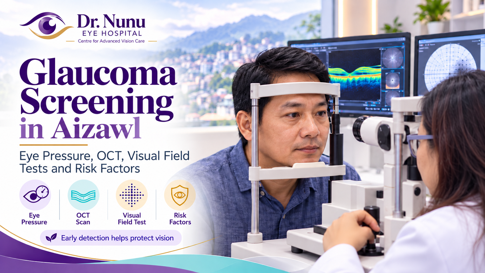
Glaucoma is a group of eye diseases that damage the optic nerve—the nerve that carries visual information from the eye to the brain. It is an important cause of irreversible vision loss because damage that has already occurred cannot usually be restored. The encouraging news is that early detection and appropriate treatment can often slow or prevent further loss of sight.
Most common forms of glaucoma develop gradually and may not cause pain or obvious symptoms in the early stages. Central vision can remain clear while peripheral, or side, vision is slowly affected. For this reason, glaucoma screening in Aizawl should be based on a comprehensive eye examination rather than waiting for symptoms.
A proper glaucoma assessment looks at several pieces of information together. Eye pressure is important, but pressure alone cannot confirm or exclude glaucoma. Examination of the optic nerve, OCT imaging, visual-field testing and assessment of individual risk factors all help the ophthalmologist decide whether the eyes are healthy, require monitoring or need treatment.
What is glaucoma?
The optic nerve contains more than a million delicate nerve fibres. Glaucoma gradually damages these fibres. In many cases, the damage is associated with pressure inside the eye, known as intraocular pressure or IOP. Eye pressure is created by a clear fluid that is continuously produced and drained from the front of the eye. When the drainage system does not work efficiently, pressure may rise.
High eye pressure increases the risk of glaucoma, but it is not the whole story. Some people have pressure above the usual range without optic-nerve damage; this is called ocular hypertension. Other people develop glaucoma even though their measured pressure is within the statistically normal range. This is often called normal-tension glaucoma. The amount of pressure an optic nerve can tolerate differs from person to person.
The two broad adult categories are open-angle glaucoma and angle-closure glaucoma. Open-angle glaucoma usually progresses slowly and silently. Angle closure occurs when the drainage angle between the iris and cornea becomes too narrow or blocked. A sudden complete blockage can cause a rapid pressure rise and is an eye emergency.
Why glaucoma screening matters
Early glaucoma is often symptom-free. A person may read well, recognise faces and work normally while gradual peripheral-vision damage is developing. The brain can also compensate for small missing areas, making the change difficult to notice without testing.
Vision already lost from glaucoma is usually permanent. Screening aims to identify disease—or a high-risk “glaucoma suspect”—before major functional vision loss occurs. If glaucoma is found, lowering eye pressure through medication, laser or surgery can reduce the risk of further damage. The treatment plan depends on the type and severity of disease and the patient’s overall circumstances.
Who should consider glaucoma screening in Aizawl?
Anyone can develop glaucoma, but some people need particular attention. A comprehensive baseline eye examination is useful from around age 40, with follow-up based on the findings and risk profile. People with significant risk factors may need earlier or more frequent checks.
- Increasing age: glaucoma becomes more common as people grow older.
- Family history: having a parent, sibling or close blood relative with glaucoma raises risk.
- Raised eye pressure: higher IOP is an important and modifiable risk factor.
- Diabetes or high blood pressure: these conditions make regular comprehensive eye care especially important.
- High short-sightedness or long-sightedness: certain refractive patterns can be associated with particular glaucoma risks.
- Thin central cornea: corneal thickness affects both risk assessment and interpretation of pressure readings.
- Previous eye injury or surgery: trauma and some eye conditions can cause secondary glaucoma.
- Long-term steroid use: steroid eye drops, tablets, inhalers, creams or injections may raise eye pressure in susceptible people.
- Inflammation inside the eye: uveitis and related disorders may increase glaucoma risk.
- Narrow drainage angles: people with anatomically narrow angles may be vulnerable to angle closure.
Having a risk factor does not mean that a person definitely has glaucoma. It means the ophthalmologist may recommend a more detailed evaluation or closer follow-up.
Why an eye-pressure check alone is not enough
Community screenings sometimes rely on a quick air-puff pressure check. This can identify some people who need further examination, but it cannot diagnose glaucoma on its own. A patient can have raised pressure without glaucoma or glaucoma with a pressure reading that appears normal.
Pressure also varies during the day and can be influenced by corneal thickness, the measurement technique and other factors. A single reading is only one point in a larger clinical picture. The most reliable assessment combines pressure measurement with evaluation of the optic nerve, retinal nerve-fibre layer, visual field and drainage angle when indicated.
Eye-pressure testing: tonometry
Tonometry measures intraocular pressure. Different instruments can be used. A non-contact tonometer sends a brief puff of air toward the cornea. Applanation tonometry gently touches the numbed surface of the eye with a small instrument and is commonly used for precise clinical measurement.
The test is quick. Anaesthetic drops may be used for contact measurement, so it should not be painful. The result is recorded in millimetres of mercury, written as mmHg. Although readings above 21 mmHg are often described as higher than the usual range, there is no single safe or unsafe number for everyone. The ophthalmologist interprets the reading alongside the health and appearance of the optic nerve.
Optic-nerve examination
The optic nerve can be examined through the pupil using specialised lenses and a slit lamp or other instruments. Dilating drops may be used to provide a clearer view. The doctor assesses the colour, shape and central cup of the optic-nerve head and looks for patterns of thinning, bleeding or asymmetry that may suggest glaucoma.
Optic nerves naturally vary in size and appearance. One examination may not always distinguish normal anatomy from early disease. Photographs and OCT scans create a baseline that can be compared with future tests. Detecting change over time is often an important part of diagnosis.
What is an OCT test for glaucoma?
Optical coherence tomography, or OCT, is a non-contact imaging test that uses light to create detailed cross-sectional images of structures at the back of the eye. For glaucoma assessment, OCT measures the retinal nerve-fibre layer around the optic nerve and may also analyse layers of the macula containing nerve cells.
The patient places their chin and forehead against the instrument and looks at a target while images are captured. The scan is quick and does not expose the eye to X-rays. OCT can help identify structural thinning that may occur before a patient notices vision loss.
An OCT colour map is not a diagnosis by itself. Scan quality can be affected by high refractive error, cataract, eye movement, segmentation errors or an optic nerve that differs from the reference database. The ophthalmologist interprets the images in combination with the clinical examination and visual-field results. Repeat scans help show whether thinning is stable or progressing.
What is a visual-field test?
A visual-field test measures central and peripheral vision. During an automated field test, one eye is covered while the other looks at a central target inside a bowl-shaped machine. Small lights appear in different locations, and the patient presses a button whenever a light is seen. The procedure is then repeated for the other eye.
Glaucoma often creates characteristic blind spots in side vision before central vision is affected. Field testing therefore measures how the optic nerve is functioning, while OCT provides structural information. The two tests are complementary.
The test requires concentration. Blinking, tiredness, looking away from the central target or pressing when no light was seen can reduce reliability. It is common for a doctor to repeat an abnormal or uncertain result. Familiarity also helps many patients perform more consistently on later tests.
Gonioscopy: checking the drainage angle
Gonioscopy examines the drainage angle where the iris meets the cornea. After numbing drops are applied, the ophthalmologist gently places a special mirrored lens on the eye. This helps determine whether the angle is open, narrow or closed and can reveal pigment, abnormal blood vessels or scarring.
Knowing the angle configuration is important because open-angle and angle-closure glaucoma require different decisions. A narrow angle does not always mean glaucoma is present, but it may require counselling, monitoring or preventive treatment depending on the individual findings.
Corneal-thickness testing: pachymetry
Pachymetry measures the thickness of the central cornea. A pressure reading can appear higher in a thicker cornea or lower in a thinner cornea than the true pressure-related risk might suggest. A thin cornea is also an independent risk factor considered when estimating whether ocular hypertension may progress to glaucoma.
Pachymetry is usually a quick, painless test. The result helps the ophthalmologist interpret IOP more meaningfully, but pressure should not be adjusted by a simple formula without considering the full examination.
When glaucoma symptoms are an emergency
Most open-angle glaucoma does not cause sudden symptoms. Acute angle closure is different and can cause a rapid, severe pressure rise. Seek emergency eye care immediately for:
- sudden severe eye pain or headache;
- a red eye with blurred or reduced vision;
- coloured halos around lights;
- nausea or vomiting with eye pain;
- a suddenly fixed or unusual-looking pupil;
- rapid loss of vision.
These symptoms should not wait for a routine appointment. Contact an eye hospital urgently. Our guide to emergency eye care in Aizawl explains other warning signs that require prompt assessment.
What does “glaucoma suspect” mean?
A glaucoma suspect is someone whose pressure, optic-nerve appearance, OCT, visual field, drainage angle or family history raises concern, but who does not yet have definite glaucoma. Some suspects never develop the disease, while others show change over time.
Monitoring may include repeat pressure checks, optic-nerve examination, photographs, OCT and visual fields. The interval depends on the level of risk. Treatment may be recommended if the estimated risk of damage is high, if pressure is at an unsafe level for that particular eye or if progressive structural or functional change is detected.
How often should glaucoma tests be repeated?
There is no single schedule for every patient. Higher-risk people without glaucoma are often advised to have comprehensive examinations every one to two years, while glaucoma suspects may need earlier review. Patients with confirmed glaucoma may require visits several times a year depending on pressure control, disease severity and evidence of progression.
The ophthalmologist may repeat OCT and visual-field tests to establish a reliable baseline and measure change. Keeping follow-up appointments is important because comparison over time can reveal progression that is not obvious from one isolated test.
If glaucoma is diagnosed, what treatments are available?
The purpose of treatment is to lower eye pressure to a level that reduces the risk of further optic-nerve damage. Options may include prescription eye drops, laser procedures or surgery. The choice depends on the type of glaucoma, drainage-angle anatomy, target pressure, stage of disease, general health and ability to use medication consistently.
Glaucoma treatment does not usually restore vision already lost. It is designed to preserve remaining sight. Patients should use medicines exactly as prescribed and should not stop drops because the eye feels normal. Report side effects and difficulties so the ophthalmologist can adjust the plan safely.
Preparing for a glaucoma screening appointment
Bring all current spectacles, a list of medicines and any previous eye records. Tell the doctor about glaucoma in the family, past eye injuries or operations, steroid use, diabetes, high blood pressure and episodes of severe eye pain or coloured halos. If dilation is planned, vision may be temporarily blurred and sensitive to light, so sunglasses and help with transport can be useful.
A full glaucoma assessment may take longer than a routine spectacle check because several tests are needed. Rest well before visual-field testing and follow the examiner’s instructions. If you feel tired or uncomfortable, let the examiner know rather than guessing at the lights.
Patients can also read about the broader tests included in a comprehensive eye check-up in Aizawl. People with diabetes should continue their scheduled diabetic retinopathy screening as well.
Frequently asked questions
Can I have glaucoma if my eye pressure is normal?
Yes. Normal-tension glaucoma causes optic-nerve damage even though measured pressure is within the usual statistical range. This is why pressure testing must be combined with optic-nerve evaluation, OCT and visual-field testing.
Does high eye pressure always mean glaucoma?
No. Some people have ocular hypertension without nerve damage or field loss. They still require individual risk assessment and monitoring because higher pressure increases the chance of developing glaucoma.
Is an OCT scan painful?
No. OCT is a quick, non-contact imaging test. The patient looks at a target while the machine captures detailed images using light.
Why might my visual-field test need to be repeated?
The test depends on concentration and response. Tiredness, fixation changes or unfamiliarity can affect the result. Repeating the test can confirm whether a suspected defect is genuine and helps monitor change over time.
Can glaucoma be cured?
Glaucoma is generally a lifelong condition rather than one that is permanently cured. Treatment and monitoring can often control pressure and slow or prevent further damage.
Where can I arrange glaucoma screening in Aizawl?
Patients in Aizawl and across Mizoram can contact Dr. Nunu Eye Hospital & Research Centre for an eye assessment. Dr. Lynapawngia Solo can review the clinical findings and advise whether monitoring, additional testing, treatment or referral is appropriate.
Book a glaucoma assessment in Aizawl
Glaucoma may be silent, but a comprehensive examination can identify important warning signs before a person notices substantial vision loss. Eye pressure, OCT, visual fields and optic-nerve examination each answer different questions, and the most reliable assessment considers them together.
If you are over 40, have a family history of glaucoma, have been told your eye pressure is high, use steroids long-term or have another risk factor, ask an ophthalmologist how often your eyes should be checked. For appointments at Dr. Nunu Eye Hospital & Research Centre, call +91 98620 19216.
This article provides general health information and does not replace a comprehensive eye examination or personalised medical advice. Test schedules and treatment decisions should be made with an ophthalmologist.
Leave a Reply